Cell viability must be determined in order to understand how cells behave and how they respond to analysis or therapeutic testing. To perform accurate cell viability tests, researchers must optimize their procedures for distinguishing healthy cells from dead or damaged cells. Our cell enrichment technologies identify and remove unwanted dead or degraded cells from a sample in a gentle and efficient manner, leaving healthy cells for further study or downstream research applications.
Cell Viability
The number of living cells in a population is referred to as cell viability. Understanding cell behavior and assessing efficacy require determining the viability of cells in a sample throughout research and treatment development. Cell viability assays enable researchers to infer the overall health of cells in samples, improve experimental procedures, and determine whether cells remain healthy after therapeutic testing.
Percentage of Cell Viability
As a measure of overall cell survivability, the cell viability percentage of a sample is a useful check of research efficacy. Calculating cell viability percentages at various stages of analysis, for example, informs decisions about which procedures are best suited for experimental success.
How To Check the Viability of Cells
Cell viability assays for cells in culture are used by researchers to determine cell health. Typically, these assays monitor cell proliferation, metabolic activity, or ATP content. Cell viability can also be calculated by first performing cell toxicity assays, which count the number of dead or damaged cells in a sample, and then subtracting these unhealthy cells from the total population to determine the number of viable cells.
Assays for Cell Proliferation
Cell proliferation refers to a cell’s ability to reproduce through cell division, also known as cytokinesis. Because only healthy cells divide and proliferate, while damaged, dying, or dead cells do not, researchers use cell proliferation assays to calculate the overall cell viability percentage by measuring the increase in cell count in a population.
Cell proliferation assays typically measure DNA synthesis or overall DNA content within replicating cells via imaging, flow cytometry, immunofluorescence, or enzyme-linked immunosorbent assays to determine the number of cells actively dividing within a culture (ELISA).
Cell cycle analyses are a type of proliferation assay that monitors how healthy cells progress through the cell cycle. Viable cells that proliferate first expand in size, then synthesize and replicate their DNA. Following DNA replication, cells grow even larger before entering mitosis, a process in which cells enclose the two replicated DNA strands with distinct nuclear membranes.
DNA synthesis assays are another method of analyzing proliferation to determine cell viability. Similar to cell cycle analyses, researchers incorporate labeling agents into freshly synthesized DNA and then monitor their concentrations over the course of the investigation. Since the amount of DNA synthesis among the cells under study is correlated with the presence of these labeling substances in succeeding cell generations, this correlation can be used to gauge how well healthy cell division proceeds in the sample population.
Assays of Metabolic Activity
Aside from actively dividing cells or cells that aren’t proliferating, metabolic activity levels in each population can also be used to infer cell viability.
MTT or XTT assays first incubate a sample with a yellow dye; mitochondrial enzymes in healthy cells then convert these dyes into a purple byproduct while producing energy to power cell activity. The amount of these byproducts in the sample can then be used to calculate the number of viable cells in a population by counting the number of purple-colored cells.
Adenosine triphosphate, or ATP, is a molecule produced during the normal cellular function that provides energy to living cells. Another type of metabolic analysis is ATP measurement, which tracks the production of ATP or the presence of byproducts of ATP consumption during normal metabolic processes.
Cell Toxicity Assays
Cell viability can also be derived from cell toxicity assessments, which are a measure of the number of cells that die or become damaged, either naturally during the cell cycle, which results in cell death, or after treatment with pharmacological agents during therapeutic development and testing.
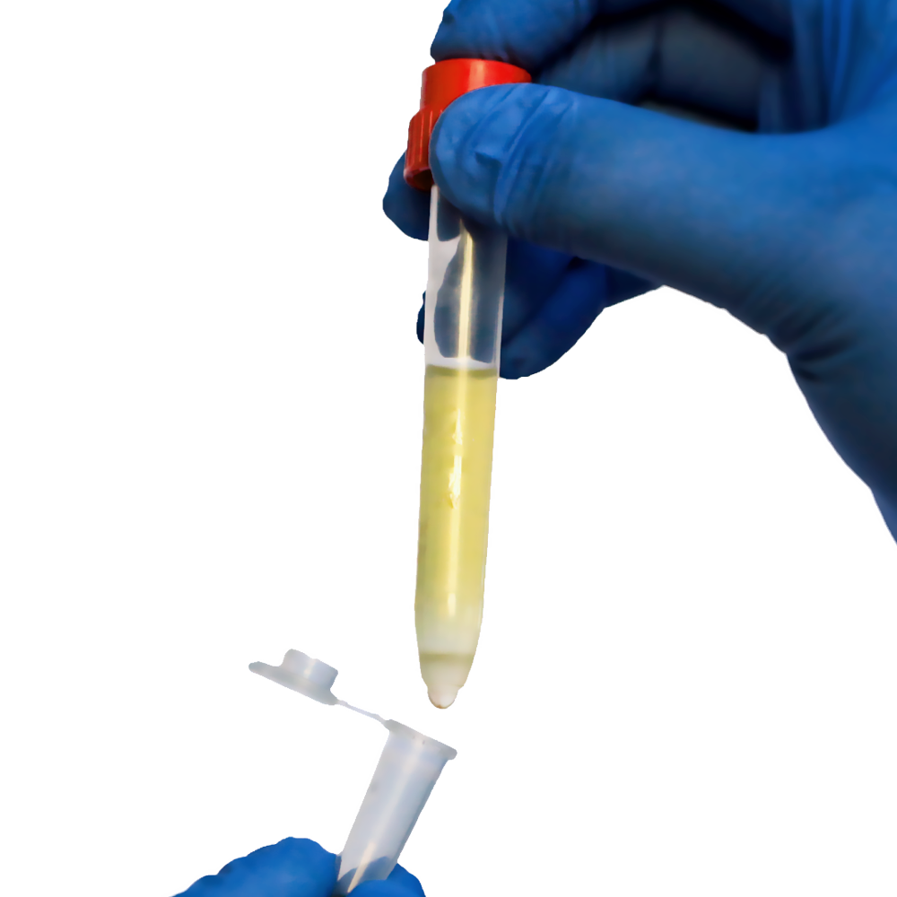
Improving Cell Viability with Pluribead
Cell degradation in a population can occur as a result of natural cell death, therapeutic testing, or experimental procedure. When cells die or are damaged, they leave behind cellular debris. Both unhealthy cells and the debris they produce can impair the accuracy of cell viability assays if they are counted as whole cells or are unable to be differentiated.
The sample population being tested must be made up of viable cells in order to determine how a particular process or treatment will affect healthy cells in the body. As a result, the ability to distinguish healthy cells from cellular debris or dead cells within a sample is critical for accurately determining cell viability and the efficacy of therapeutic testing.
Pluriselect has created a breakthrough technology for healthy cell separation from dead or damaged cells that is inexpensive, simple to use, and produces viable cell populations of high purity.
When the specific antibody binds directly to the cells using positive selection, all undesirable cells are separated from the labeled and desired cells during the subsequent enrichment steps because they are all unbound. When coupled to a solid phase, a cell strainer is the simplest way to keep labeled cells in place. Any type of sample material, such as PBMC, secretion or excretion material, whole blood, buffy coat, spleen, liver, and so on, may be used.
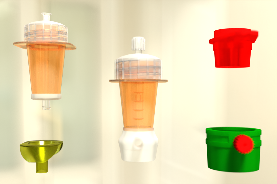
Two Different Bead Sizes are Available
S-pluriBead: It is used in large sample volumes with a small number of targets.
M-pluriBead: A versatile material that can be used to achieve multiple goals while using less material.
Key features of Pluribead
- No Sample Preparation: Use a sample volume ranging from 200 l to 45 ml with no erythrolysis, gradient centrifugation, or other techniques.
- Any type of sample material, such as PBMC, secretion/excretion material, brain homogenate, spleen, liver, buffy coat, whole blood, and so on, can be used.
- Suitable for Isolation from a Wide Range of Species: Isolate from sheep, mice, rats, cows, dogs, and other animals.
- Cell division can begin within five minutes of isolation.
- PluriBead Cascade Simultaneous Cell Isolation: Separate two different cell types from the same sample material at the same time.
- Using sequential cell isolation, you can isolate up to six different targets from a single sample.
To learn more about the importance of cell viability and how Pluribead is revolutionizing cell enrichment and cell viability assays, you can visit our website or talk to our support team.
Reference:
NCBI
Science Direct
Nature
 English
 French
 German
 Spanish
 Belgium
 Italian
 Brazil
 Chinese Mandarin