Circulating tumor cells (CTCs) in the bloodstream play an important role in metastasis formation. Know more about the fast and effective tool for cell separation.
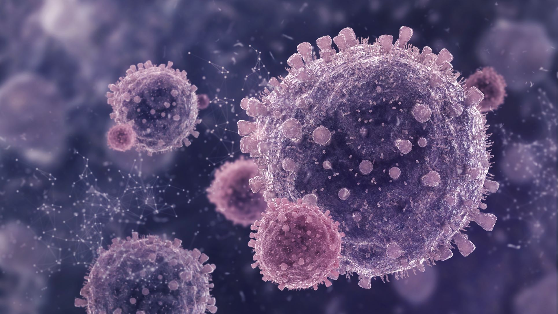
CTCs are a major contributor to cancer metastases and plays for the enumeration of CTCs from cancer patients’ whole blood with high capture and detection efficiency. Existing and ongoing research on Circulating Tumor Cells separation methods has revealed cell characteristics that are useful in cancer monitoring and treatment design.
CTCs are highly valued in medical research because they can alert doctors to the presence of a tumor before imaging. Working backward, scientists can use CTCs to determine the approximate size and location of a primary mass in order to begin treatment as soon as possible.
The most significant limitation of CTC isolation is its scarcity. In order to increase the effectiveness of circulating tumor cell separation and enumeration, numerous techniques have been created and are continuously being improved. They have proven to be capable of not only identifying tumors but also giving us their essential details.
CTC Separation
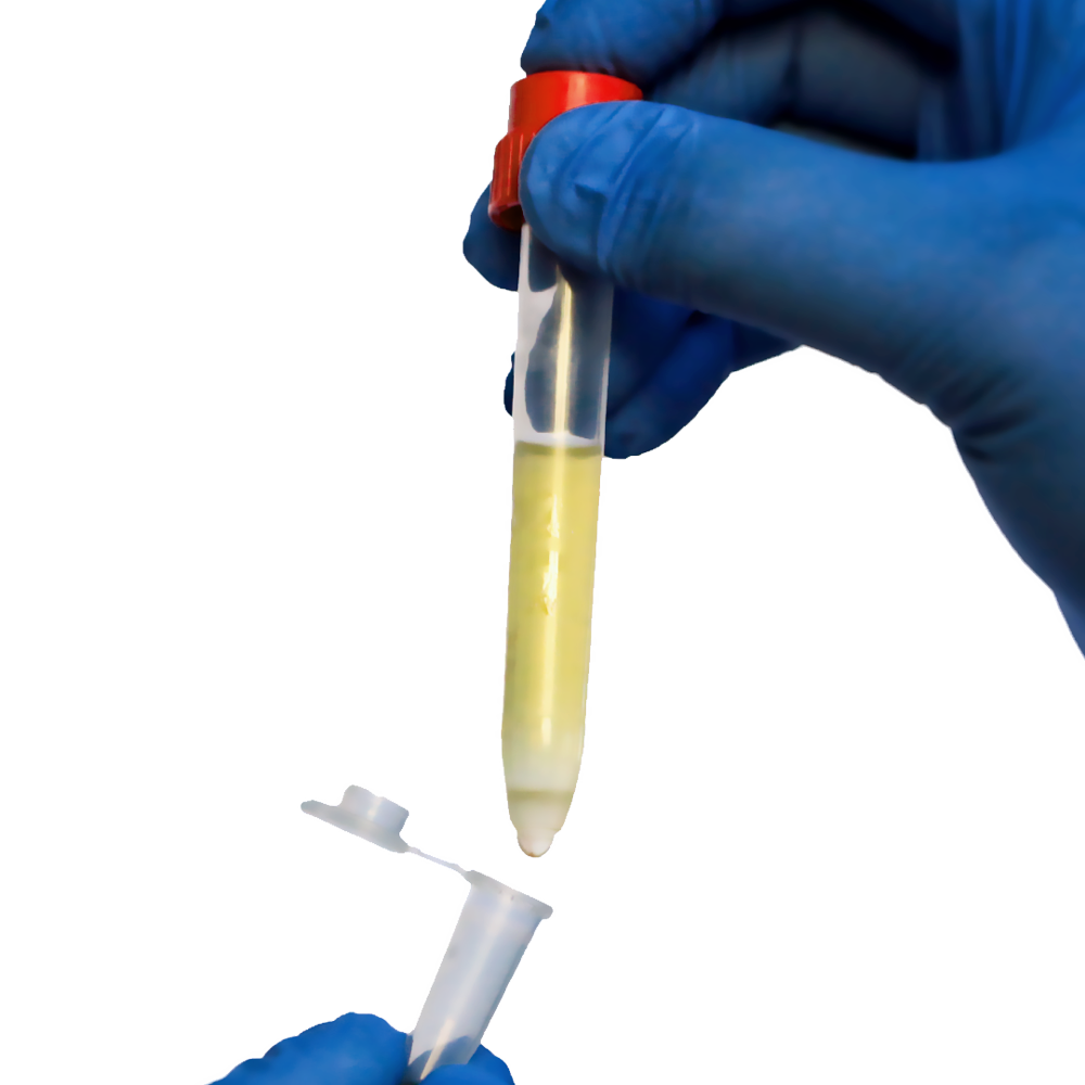
The process of isolating CTCs from residual blood cells is known as CTC separation. Red blood cells (RBCs), white blood cells (WBCs), and other substances can all impair the clarity of subsequent experimental results.
CTCs must be isolated and purified before they can be studied properly. CTC Enrichment refers to the process of isolating and purifying CTCs.
CTC Process
The most popular technique for CTC separation is the use of antibodies that focus on the tumor cell surface antigens. Immune system molecules called major histocompatibility complex (MHC) molecules have the ability to recognize unidentified cells and bind peptide chains to them, enabling other cells to destroy them. To distinguish CTCs from other cells, this process can be artificially replicated.
This can be difficult because CTCs exhibit varying levels of epithelial markers and different antigens can form on similar cells. As a result, they are more difficult to retrieve or mark with antibodies. They also risk being destroyed by the immune system before being extracted.
When attempting to separate by size or density, CTC uses blood filtration and Density Gradient Centrifugation. Filtration has a high separation efficiency and is easily automated, but it can become inaccurate due to cell accumulation and unwanted cells squeezing through filters. Density gradient centrifugation aids in the grouping of CTCs but leaves them heavily contaminated by RBCs.
Limitations
CTCs are not completely saturated in blood samples. These cells are extremely difficult to isolate due to their scarcity. Many current methods struggle to purify the sample without the use of additional steps. Even after cleaning with a density gradient and microfluidics, a sample can still contain more than 90% RBCs.
When sorting, these RBCs take up space that would normally be filled by more CTCs; they also make it difficult to culture rare cells due to the physical space they take up. The presence of these cells contaminates enriched CTC samples, making it more difficult to secure results for downstream applications.
A New Approach To Colon Carcinoma Circulating Tumor Cell (CTC) Detection Using Pluribead
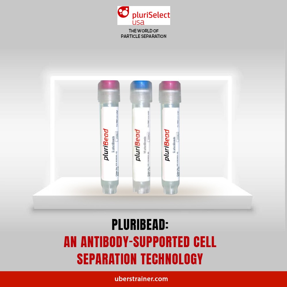
We present a novel method for screening colon carcinoma circulating tumor cells (CTCs) with pluriBead containing a tumor-associated EpCAM antibody.
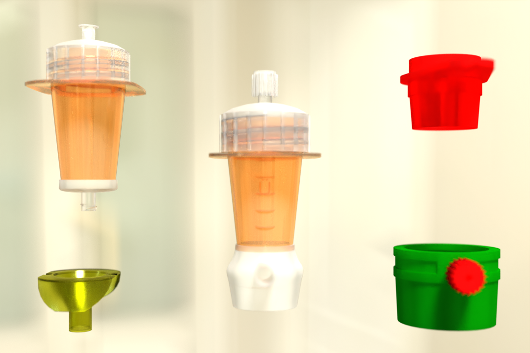
This method is based on non-magnetic cell separation technology. There is no need for sample preparation. EpCAM-pluriBead® can be injected directly into whole blood. The method can also be used to separate single cells from various biological fluids. The sample volume can also be increased to increase its sensitivity.
The bound EpCAM-positive colon carcinoma cells can be easily included after molecular-genetic experiments to determine their mutation status. K-ras mutation status is a tool for predicting response to anticancer therapy in the case of colon carcinoma. The new technique thus serves as a quick and reliable tool for early cancer detection.
Steps Involved In Tumor Study
- Labeling
Add EpCAM-pluriBead® to your sample
- Mixing
30 minutes of gentle incubation (recommended with pluriPlix®).
- Isolation
Captured target cells are separated using appropriate sieves.
- Washing
Use wash buffer to clean the sieve. Lyse cells with Trizol®.
- CTC Processing
Approaches for the study of cancer cells:
– RNA/DNA isolation
– Cell culture experiments
Conclusions And Perspectives
Using EpCAM-pluriBead®, we developed a new method for detecting circulating colon tumor cells in the blood. The captured cells can therefore be used for additional molecular-genetic screening for particular tumor-related markers. The developed “in situ immunoblasts pcr” method eliminates the need for pre-isolated RNA/DNA and can significantly shorten analysis time. Finally, the goal of this work is to improve the method’s sensitivity by increasing sample volume, changing cell tumor lines, and/or changing antibody specificity.
Check Our Cell Separation Technology Today
Regarding cell isolation and separation needs, we have you covered. Look no further than Pluribead if you’re looking for a method to increase the quality and viability of sorted cells while significantly cutting time and costs.
Despite the fact that research on these disseminated tumor cells has accelerated significantly, it has been constrained by their scarcity. We anticipate the development of cell separation and enrichment fusion techniques that will aid in the prevention of cell loss.
A variety of specific markers is also likely to improve enrichment results, as cells that can avoid EpCAM selection may also be captured. Given their enormous potential to help change the enigmatic situation of solid tumors, we can conclude that CTCs will undoubtedly become an unavoidable part of solid tumor malignancies in the near future.
Please contact us if you have any questions, and try our products as soon as possible.
Reference:
Nature
Science Direct
NCBI
 English
 French
 German
 Spanish
 Belgium
 Italian
 Brazil
 Chinese Mandarin


