Dendritic cells (DC) are antigen-presenting cells (APCs) that are essential components of the adaptive immune system. We will talk about how Pluribeads can help with dendritic cell separation.
Dendritic cells (DC) are thought to be the most effective antigen presenting cells (APC), with the unique capacity to start, coordinate, and control adaptive immune responses. They were first discovered in the mouse spleen due to their peculiar shape and ability to activate naive lymphocytes.
Although their capacity to capture, process, and present antigens is thought to be their primary characteristic, they exhibit striking phenotypic heterogeneity and their actions can have a variety of effects.
Dendritic cells (DCs), so called because of their probing, “tree-like” or dendritic shapes, are in charge of the start of adaptive immune responses and serve as the immune system’s “sentinels.” In 1868, Paul Langerhans described DCs in human skin but mistook them for cutaneous nerve cells. DCs are leukocytes derived from bone marrow (BM) and are the most potent type of antigen-presenting cells.
They can also be grown in vitro from BM and blood using different combinations of growth factors, such as GM-CSF and Flt3 ligands.
We will talk about Pluribead cascade straining that will aid in the separation of Dendritic cells in this blog.
DCs are trained to capture and process antigens, converting proteins into peptides that are then presented on MHC molecules recognized by T cells. Although all DCs are capable of antigen uptake, processing, and presentation to naive T cells, DC subtypes have distinct markers and differ in location, migratory pathways, detailed immunological function, and reliance on infections or inflammatory stimuli for their generation.
The phenotype and function of DCs play an extremely important role in initiating tolerance, memory, and polarised T-helper 1 (Th1), Th2, and Th17 differentiation during the development of an adaptive immune response.
Dcs Linking Innate And Adaptive Immunity
Because DCs have numerous cytoplasmic processes, they have a large surface area that allows them to interact with a large number of surrounding cells, such as T cells, natural killer cells, neutrophils, epithelial cells, and so on. In experiments, for example, only one mature DC (mDC) is needed to stimulate 100-3000 T cells.
DC precursors migrate from the BM through the bloodstream to almost every non-lymphoid tissue, where they remain in an immature state (iDC), sampling their environment continuously via endocytosis, macropinocytosis, and phagocytosis. They can extend their processes through epithelial tight junctions to increase antigen capture even when there is no obvious infection/inflammation.
During pathogen invasion, resident iDCs detect intruders using pattern recognition receptors (e.g., TLRs), capture antigens and exit the tissue quickly. In response to chemokines such as CCL19 and CCL21, they crawl through cells, cross the endothelium of lymphatic vessels, and migrate to draining lymph nodes (LN). DCs mature phenotypically and functionally as they migrate from peripheral tissues.
Most notably, they cease antigen capture while increasing the expression of co-stimulatory molecules like CD80 and CD86, as well as the chemokine receptor CCR7, and secrete pro-inflammatory cytokines like TNF- and IL-12. DCs migrate to T-cell zones after reaching the LN’s subcapsular sinus. Interdigitating DCs are actively involved in the presentation of antigens to T cells in this case.
Subpopulations of human dendritic cells and monocyte-derived dendritic cells
There are two types of dendritic cells: migratory non-lymphoid tissue DC and resident lymphoid tissue DC. Both are diverse cell populations that can be divided into distinct subsets based on their genetic makeup and phenotypic markers. The discovery that some mouse resident splenic and thymic DCs expressed CD8, but not all of them, led to the first identification of various DC subsets.
Despite recent data characterizing DC subpopulations in the human lung and intestine, the identification of human DC subpopulations is not as advanced as that of mouse DC, largely because human tissues are not as accessible. Instead, most studies on human DC have been conducted in peripheral blood or skin.
In an effort to compare clearly defined murine subpopulations with those found in human peripheral blood, recent efforts have been made to understand the ontogeny and function of human DC subsets. A myeloid (MP) and lymphoid (LP) precursor, a CD34+ hematopoietic precursor, is the source of DC. Monocyte and common DC precursors are produced when MP differentiate into monocyte, macrophage, and DC precursors (MDP) (CDP). CDP has the ability to differentiate into preclassical DC or plasmacytoid DC (pDC) (pre-cDC). The two main cDC subpopulations, cDC1 and cDC2, descended from pre-cDC. The identification of DC subset precursors in peripheral blood and improved characterization of DC ontogeny made possible by recent technologies, like single cell RNAseq, show that the commitment to a DC subset may occur early in both mice and humans.
Pluribeads For Dendritic Cells Separation
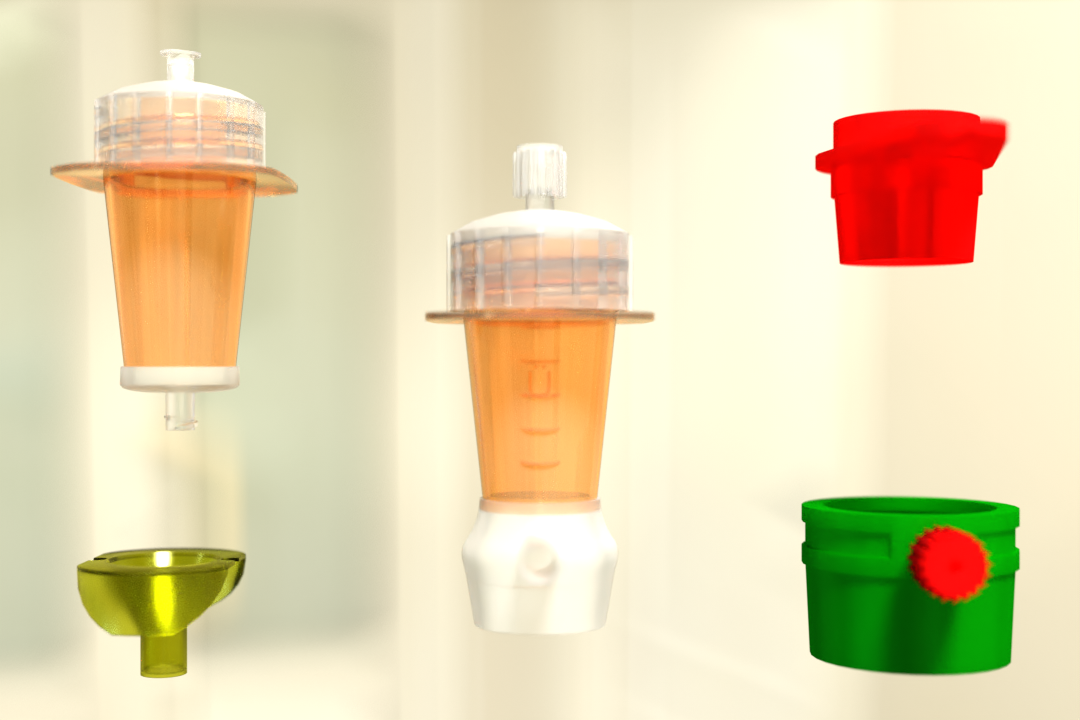
Pluribead is one such antibody cell separation technology that helps in the gentle and safe isolation of Dendritic Cells and operates without the use of any magnetic components.
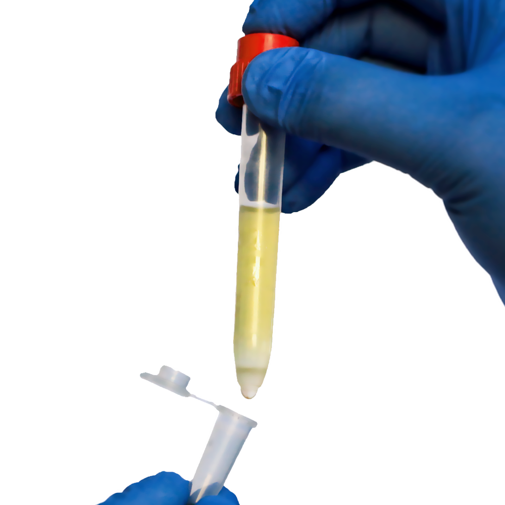
The method is simple: your pluriBeads (which contain bound target cells) are sieved through a strainer, with your target cells remaining on top and unwanted cells passing through. You are now ready to proceed with your target cells after detaching.
Two Different Bead Sizes are Available
- S-pluriBead: This type of bead is used in large sample volumes with a limited number of targets.
- M-pluriBead: A versatile material that can be used to accomplish multiple goals with less material.
Key features of Pluribead
- Use a sample volume ranging from 200 l to 45 ml with no erythrolysis, gradient centrifugation, or other techniques.
- Any type of sample material, such as PBMC, secretion/excretion material, liver, spleen, buffy coat, brain homogenate, whole blood, and so on, can be used.
- Isolate from a Variety of Species: Isolate from sheep, mice, rats, cows, dogs, and other animals.
- PluriBead Cascade Straining for Simultaneous Cell Isolation: Separate two different cell types from the same sample material at the same time.
- You can isolate up to six different targets from a single sample using sequential cell isolation.
You can see the difference for yourself by using our products. Order our cell separation products right away, and don’t forget to look over our Programs and Special Offers!
Refernce:
News Medical
Frontiersin
NCBI
 English
 French
 German
 Spanish
 Belgium
 Italian
 Brazil
 Chinese Mandarin