We’ll discuss buffy coat preparation and how it’s made from whole blood in this blog. Additionally, we will discover how Pluribead and Plurispin support this procedure.
Millions of cells are suspended in a liquid called plasma to form blood. Red blood cells (which carry oxygen), white blood cells (which fight infection), and platelets are examples of these cells (which help with clotting). Examining red blood cells is simple due to their abundance; thousands of red blood cells can be seen in a single drop of blood. White blood cells, on the other hand, are present in very small numbers and are more difficult to examine.
Examining a buffy coat smear is the quickest way to examine large numbers of white blood cells. The buffy coat is simply a collection of all of the white blood cells and platelets in a blood sample.
We will also find out how Plurispin Antibody Cell Separation technology is used in buffy coat extraction.
Importance of a Buffy Coat
The immune system of humans is built on white blood cells. Diverse types work together to identify foreign objects, get rid of health risks, and build a durable defense against infections. The first responders of the human body are platelets, which rush to afflicted areas and halt blood flow by adhesion. In the buffy coat, WBCs and platelets are both present in large quantities. Due to its high concentration of WBCs and platelets, the buffy coat is a vital bio-fluid in the field of medical research. This allows academic professionals to conduct experiments and study these vital cells and how they function in the body — which can shape the direction of medicine and effective patient treatment.
The buffy coat has uses in disease testing and treatment in addition to its research implications. The buffy coat can be used as a screening tool to check for the presence of dangerous diseases like malaria because important immune cells congregate in this layer. Additionally, by further purifying them, platelets can be used therapeutically to boost counts in patients with low platelet counts.
Buffy Coat Cells
There are four different types of cells that make up a buffy coat:
- Lymphocytes are a particular type of WBC that contribute significantly to human immunity because they produce antibodies. T lymphocytes, B lymphocytes, and natural killer cells are further divided into lymphocytes.
- White blood cells called monocytes help the immune system response by securing and consuming harmful microorganisms.
- Granulocytes are an immune cell type that is further divided into neutrophils, basophils, and eosinophils. In that order, they secrete chemicals, engage in parasite attacks, and kill bacteria and fungi.
- Platelets – Also referred to as thrombocytes, these cells are not RBCs or WBCs. They are minuscule, colorless cell fragments that travel throughout the body, clotting when necessary to stop bleeding.
Humans rely on the buffy coat’s cells for both disease defense and wound healing.
How should fresh whole blood be prepared for a Buffy Coat fraction?
Even though a buffy coat’s components are present in whole blood, they do not aggregate until centrifugation takes place. Prior to centrifugation, researchers should take a number of safety measures to guarantee the extraction procedure is successful.
There are two ways to get a buffy coat ready. One way is by centrifuging whole blood donations to separate red blood cells, plasma, and buffy coats. The second option is to filter the blood to remove leukocytes.
Preparation of buffy coat from scratch
Making your own buffy coat is an alternative. Therefore, simply adhere to the following succinct protocol:
- Mix one part washing buffer with one part whole blood.
- While the brake is off, centrifuge the diluted whole blood for 10 minutes at 200 x g.
- Remove the interphase leukocytes (buffy coat)
A whole blood sample should always be handled with the same caution as if it were possible that it could transmit pathogens that cause infectious diseases. The time has come to separate the buffy coat from a whole blood sample after all the necessary preparations have been made.
Buffy Coat Extraction
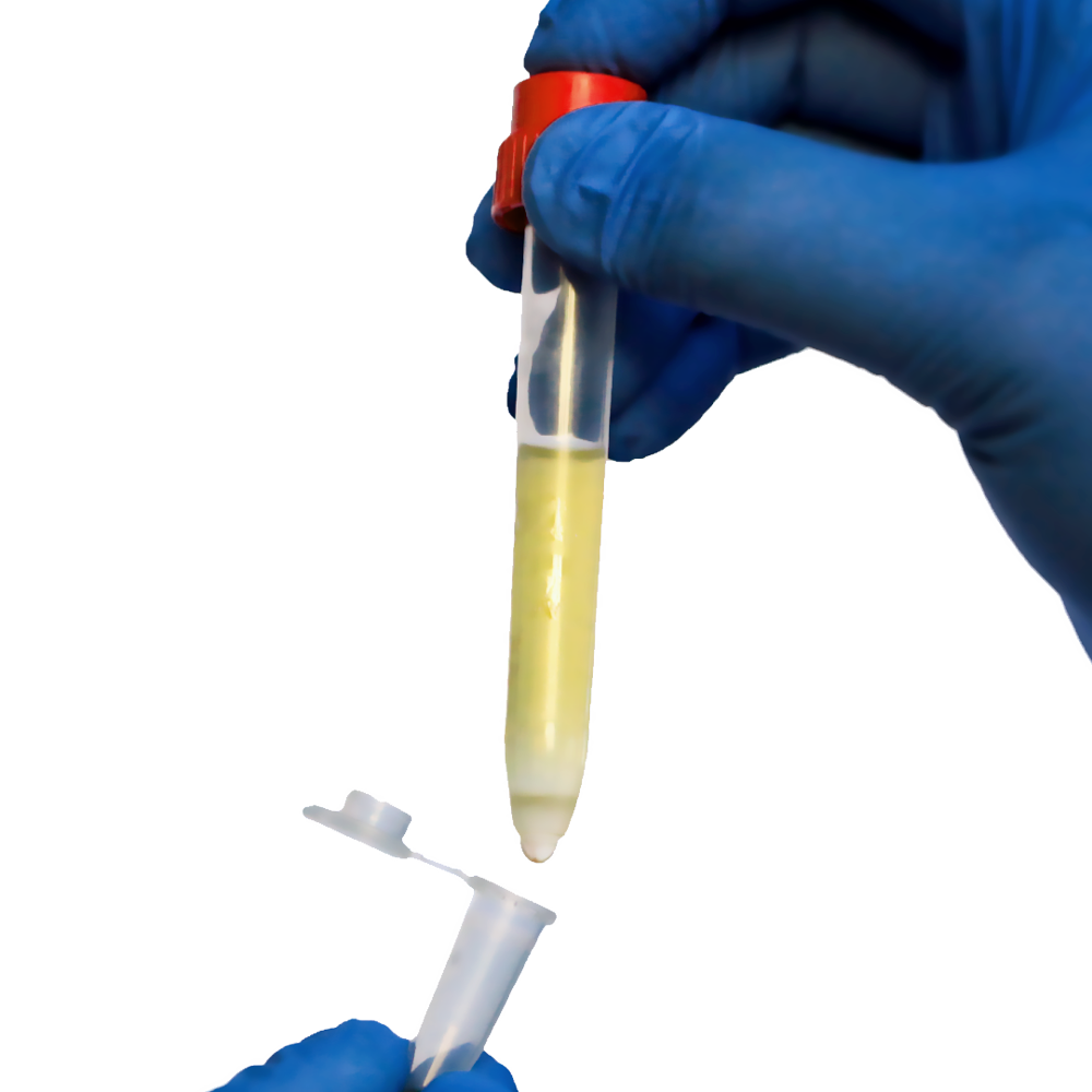
The buffy coat is separated from the plasma and RBC by centrifuging the prepared whole blood sample. After centrifugation, there will be a thin layer between the RBC and plasma that accounts for about 1% of the sorted sample – the buffy coat. The experimenter uses a tiny pipette to gather and transfer the buffy coat to a different container.
The buffy coat is in direct contact with isolated RBCs, while PBMCs are isolated from the other particles in the sorted sample by density gradient centrifugation. Using a manual extraction technique like pipetting increases the possibility that contaminating RBCs will be present in the buffy coat sample because of the size and abundance of RBCs (Buffy coats are referred to as “pink” before further processing because of this).
Researchers frequently combine centrifugation with another cell separation technique, like BACS, to get rid of any remaining RBC contamination in order to enrich the buffy coat even more.
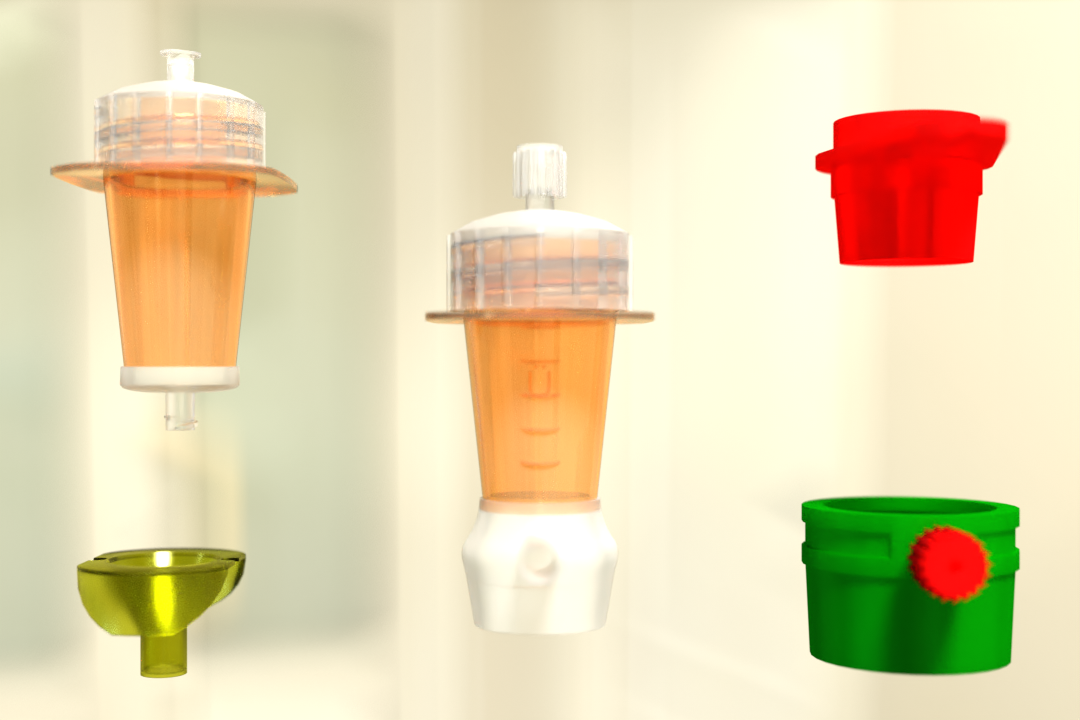
How can Plurispin be useful?
A brand-new technique for isolating negative cells from whole blood, buffy coat, or cord blood is called the PluriSpin system. This new technique isolates live, unaltered, and highly purified cells in a single step without the use of magnets or a column. As a result, there is less chance of activating or harming the target cells.
How PluriBead can help?
The unique PluriBead antibody cell separation technology does not use any magnetic components. The process is simple: Your pluriBeads are sieved through a strainer with your bound target cells on top, while the unwanted cells pass through. After detaching, you have your target cells ready.
For more information on buffy coat preparation, you can visit our website. Utilize our products to see the difference for yourself. Get started with our cell separation products today!
Reference:
Vachospitals
Science Direct
 English
 French
 German
 Spanish
 Belgium
 Italian
 Brazil
 Chinese Mandarin