Specialized precursor cells called megakaryocytes produce and release platelets into the bloodstream. Let’s explore the most efficient technologies for megakaryocyte separation.
Megakaryocytes are the cells which are present in the bone marrow that produce the platelets required for blood clotting. How megakaryocytes get to be so big and have so many nuclei is one of their mysteries. Megakaryocytes expand in size due to endomitosis, a process in which the DNA within the cell replicates numerous times without the cell dividing.
Before eventually becoming the biological equivalent of a supernova and undergoing profound changes to fragment into the thousands of platelets required for typical blood clotting, a megakaryoblastic can house more than 120 sets of nuclear DNA.
This blog discusses the best Cell Separation technologies that will help to improve workflow results.
What is the Function of Megakaryocytes?
Hematopoietic cells called megakaryocytes are in charge of making blood platelets. According to the conventional understanding of megakaryopoiesis, a mature megakaryocyte develops from hematopoietic stem cells after passing through a hierarchy of progenitor cells.
A variety of unique cytokines, chemokines, and other mediators can be produced by megakaryocytes. High concentrations of TGF- and PF4 from megakaryocytes, which are involved in the control of hematopoietic stem cells, are found in the bone marrow niche. Megakaryocytes can control neutrophil exocytosis from bone marrow by expressing the neutrophil chemoattractants CXCL1 and CXCL2 (human IL-8 equivalents).
In actuality, thrombopoietin administration can increase the number of circulating neutrophils because megakaryocytes release CXCL1/2 in response to thrombopoietin produced by G-CSF-sensing cells in the marrow stroma, which is how granulocyte colony-stimulating factor (G-CSF) mediates the release of neutrophils from the murine marrow.
Additionally, IL-1 and IL-1 can be produced by megakaryocytes as free cytokines or as microparticles. A potential role for megakaryocytes in IL-1-driven systemic inflammatory disease is established by the finding that engraftment of WT but not IL-1-deficient megakaryocytes restore the susceptibility of some disease-resistant mice to arthritis, even without the involvement of their platelets.
The only job of megakaryocytes, which are highly specialized precursor cells, is to produce and release platelets into the bloodstream. Hematologists have been fascinated by the processes by which megakaryocytes develop and give rise to platelets for more than a century. One needs high quality Antibody Cell Separation technologies to perform a Megakaryocytes separation. These cells are derived from pluripotent stem cells and go through endomitosis, a special process that allows for multiple DNA replications without cell division.
Following endomitosis, polyploid megakaryocytes enter a phase of rapid cytoplasmic expansion during which an intricate demarcation membrane system (DMS) is formed and cytoplasmic proteins and granules that are crucial for platelet function are accumulated. The cytoplasm of the megakaryocyte undergoes a significant reorganization during the last stages of development to form proplatelets, which are beaded extensions of the cytoplasm.
The Megakaryocyte As An Inflammatory Cell
Megakaryocytes have less evidence supporting their involvement in systemic inflammation than platelets do. Megakaryocytes, like platelets, can express a number of TLRs, such as TLRs 1, 2, 3, 4, and 6. Murine lung megakaryocytes express 92–97 TLR5 mRNA transcripts.
Although the functional effects of activation through these receptors are unclear, they may accelerate megakaryocyte maturation and platelet production. Like human platelets, human megakaryocytes have the ability to express the FcRIIA low-affinity IgG receptor.
Because noncanonical ligands like C-reactive protein can stimulate the high-affinity receptor FcRI, murine studies suggest that stimulation via Fc receptors may enhance the release of megakaryocyte microparticles. Since human platelets but not megakaryocytes express this receptor at the cell surface, the IgE receptor FcRI is also expressed but primarily serves to pass this receptor to platelets. It has not been determined how these receptors affect megakaryocyte function in vivo.
Megakaryocytes may also aid in the development of adaptive immunity. Megakaryocytes, like platelets, express CD40L. Megakaryocytes support plasma cell survival in the marrow environment, possibly by IL-6 and a ligand that induces proliferation. In a murine experimental model, megakaryocytes can take up exogenous antigen and cross-present it in the context of class I MHC, resulting in immune thrombocytopenia.
A variety of unique cytokines, chemokines, and other mediators can be produced by megakaryocytes. High concentrations of TGF- and PF4 from megakaryocytes, which are involved in the control of hematopoietic stem cells, are found in the bone marrow niche. 107–109 Megakaryocytes can control neutrophil exocytosis from bone marrow by expressing the neutrophil chemoattractants CXCL1 and CXCL2 (human IL-8 equivalents).
In actuality, thrombopoietin administration can increase the number of circulating neutrophils because megakaryocytes release CXCL1/2 in response to thrombopoietin produced by G-CSF-sensing cells in the marrow stroma, which is how granulocyte colony-stimulating factor (G-CSF) mediates the release of neutrophils from the murine marrow.
Additionally, IL-1 and IL-1 can be produced by megakaryocytes as free cytokines or as microparticles. A potential role for megakaryocytes in IL-1-driven systemic inflammatory disease is established by the finding that engraftment of WT but not IL-1-deficient megakaryocytes restore the susceptibility of some disease-resistant mice to arthritis, even without the involvement of their platelets.
Pluribeads: The Modern Antibody Cell Separation technology
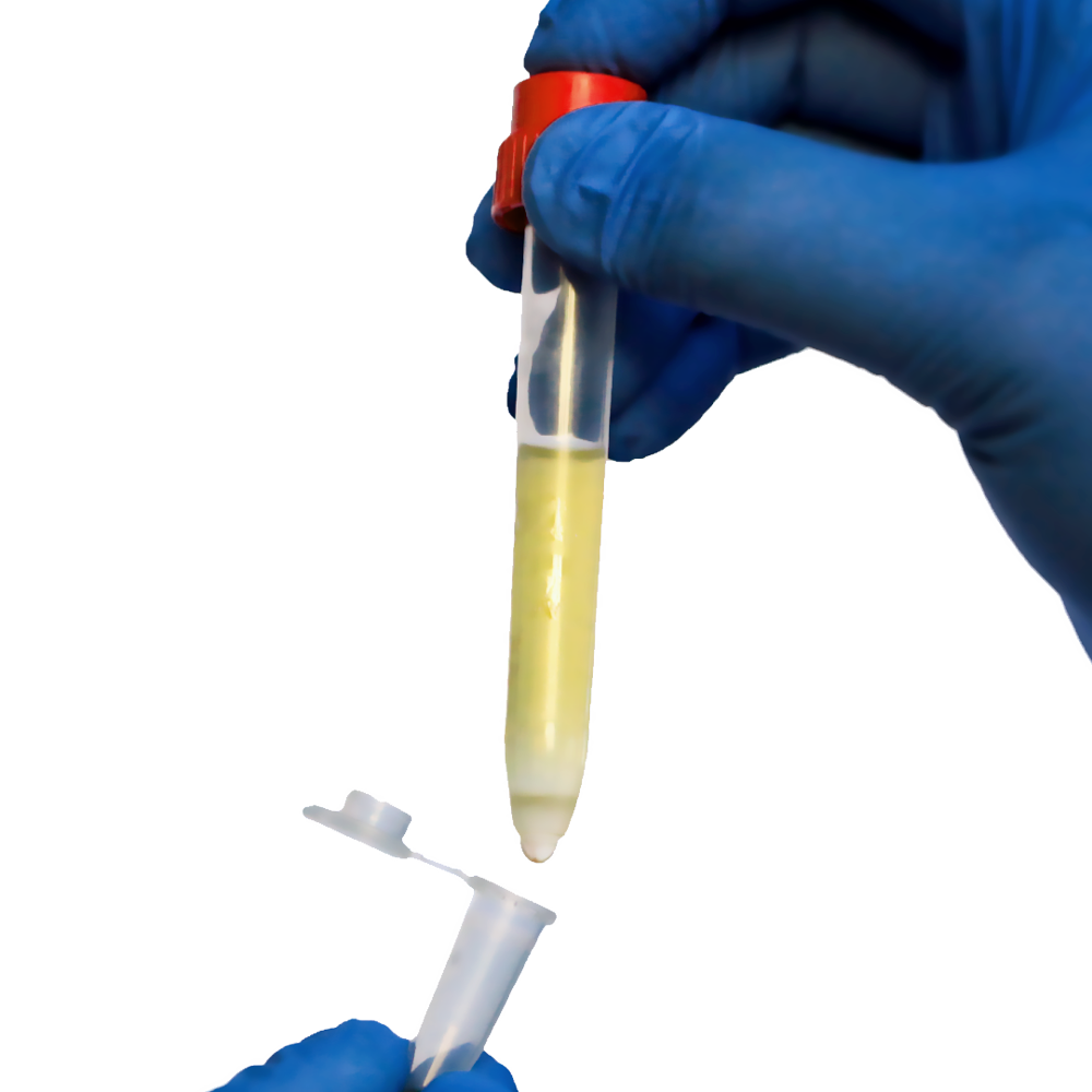
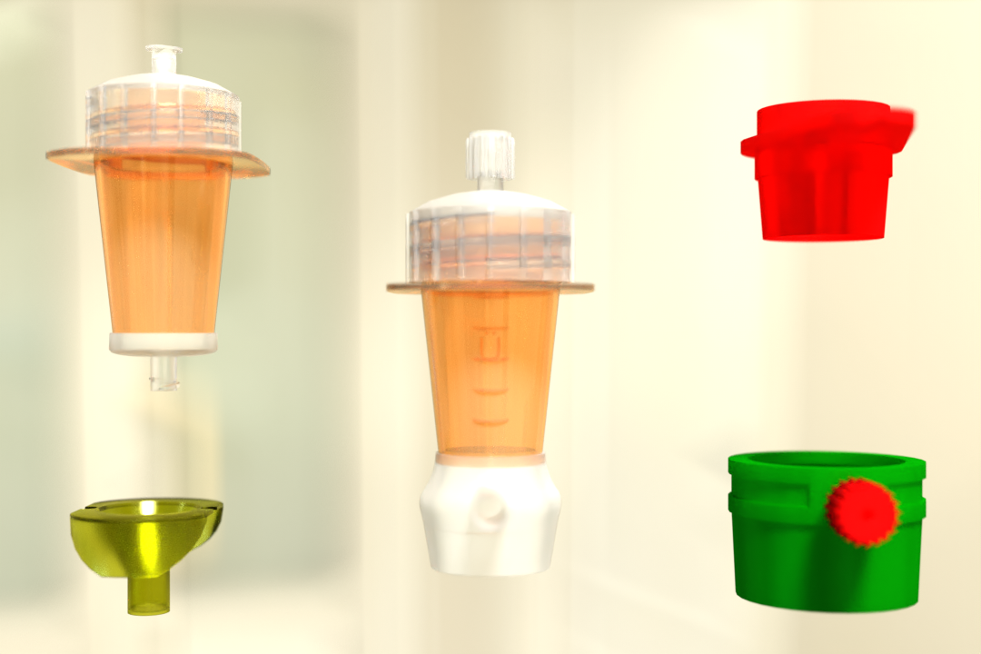
Pluribead is one such Cell Separation technology that helps in the gentle and safe isolation of Megakaryocytes. Unusual cell separation technology called PluriBead operates devoid of any magnetic components. The process is easy: Your pluriBeads (which contain bound target cells) are sieved through a strainer; the pluriBeads containing your target cells remain on top while the unwanted cells pass through. You are prepared with your target cells after detaching.
Two Different Bead Sizes Available
- S-pluriBead: It is used for a small number of targets in a large sample volume.
- M-pluriBead: A versatile material that can be used for many targets while using less material (e.g. buffy coat).
You can find out more about us by going to our website. By using our products, you can see the difference for yourself. Begin using our cell separation products right away!
Reference:
Ahajournals
Science Direct
 English
 French
 German
 Spanish
 Belgium
 Italian
 Brazil
 Chinese Mandarin