Endothelial cells play an important role in and regulate inflammatory responses. To learn more about Endothelial cells and the efficient separation technologies used to separate them, keep reading.
Endothelial cells are the cells that form the barrier between vessels and tissues. They regulate the movement of substances and fluids into and out of tissues. An impaired function can result in serious health problems throughout the body. Endothelial cells are found only in vascularized tissue and line blood vessels and lymphatic vessels. In this blog, we will explore the best Cell enrichment technologies that help to isolate Endothelial cells.
Morphology of Endothelial cells
Endothelial cells, on the other hand, come in a variety of subtypes. Endothelial cells found in lymphatic vessels are distinguished from those found in blood vessels by specific molecular markers. Endothelial cells derived from various organs add another layer of complexity. You must specify the type of endothelial cell if you want to describe its size.
Functions of Endothelial cells
Endothelial cells are no longer regarded as a passive barrier in medical research, but rather as a tissue with multiple functions. These include a variety of processes such as blood vessel formation, coagulation and fibrinolysis, vascular tone regulation, and a role in inflammation. Endothelial cells are derived from the mesoderm, a germinal layer that forms during gastrulation, an early stage of embryonic development.
Where can you find endothelial cells?
Endothelial cells can be found in all large vessels, including arteries, veins, and capillaries. An artery is made up of three layers: It is sheathed on the outside with Tunica externa, a type of connective tissue. The Tunica media is made up of smooth muscle cells, while the Tunica intima is made up of endothelial cells and lines the lumen of the blood vessel. Endothelial cells all have the same molecular characteristics: they are positive for von Willebrand factor (vWF) and CD31 glycoprotein, but negative for smooth muscle alpha-actin.
This also applies to capillary endothelial cells. Endothelial cells play a critical role in the exchange of substances such as oxygen, water, and lipids, as well as the transfer of carbon dioxide and urea from a tissue to a vessel and vice versa. Endothelial cells can differ depending on the tissue and organ.
Blood Vessels and Endothelial Cells
We now move on from tissues derived from embryonic ectoderm and endoderm to those derived from mesoderm. This middle layer of cells, located between the ectoderm and the endoderm, grows and diversifies to provide a variety of supportive functions. It gives rise to connective tissues, blood cells, and blood vessels, as well as muscle, the kidney, and a variety of other structures and cell types. We’ll start with blood vessels.
Almost all tissues rely on a blood supply, which is dependent on endothelial cells, which form the linings of blood vessels. Endothelial cells have an amazing ability to adjust their number and arrangement to meet local needs. They create an adaptable life-support system that spreads through cell migration into almost every region of the body.
Tissue growth and repair would be impossible without endothelial cells extending and remodeling the blood vessel network. Cancerous tissue, like normal tissue, requires a blood supply, which has fueled interest in endothelial cell biology. It is hoped that by inhibiting the formation of new blood vessels with drugs that act on endothelial cells, tumor growth can be slowed.
Thus, endothelial cells line the entire vascular system, from the heart to the smallest capillary, and control the passage of materials into and out of the bloodstream, as well as the transit of white blood cells. A study of the embryo also reveals that arteries and veins develop from small vessels made entirely of endothelial cells and a basal lamina: pericytes, connective tissue, and smooth muscle are added later as needed, under the influence of endothelial cell signals.
Pericyte recruitment is particularly dependent on PDGF-B secreted by endothelial cells, and in mutants lacking this signal protein or its receptor, pericytes are absent in many regions. As a result, embryonic blood vessels develop microaneurysms, which eventually rupture, as well as other abnormalities, demonstrating the importance of signals exchanged in both directions between pericytes and endothelial cells.
Arterioles and veins are the largest blood vessels, with a thick, tough wall of connective tissue and many layers of smooth muscle cells. The endothelium, an extremely thin single sheet of endothelial cells, lines the wall, separated from the surrounding outer layers by a basal lamina. The amount of connective tissue and smooth muscle in the vessel wall varies with the diameter and function of the vessel, but the endothelial lining is always present. The walls of the vascular tree’s finest branches, the capillaries, and sinusoids, are made up entirely of endothelial cells and the basal lamina, with a few scattered (but functionally important) pericytes. These are connective-tissue cells related to vascular smooth muscle cells that wrap themselves around the small vessels.
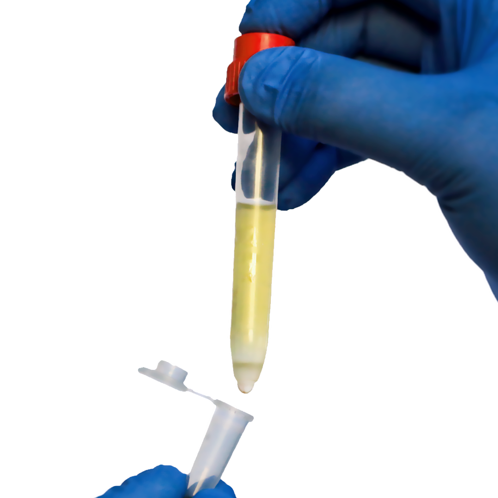
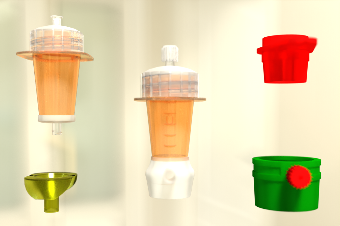
Pluribeads: an innovative approach to cell separation technology
Pluribead is one such Cell Separation technology that helps in the gentle and safe isolation of endothelial cells. Unusual cell separation technology called PluriBead operates devoid of any magnetic components. The process is easy: Your pluriBeads (which contain bound target cells) are sieved through a strainer; the pluriBeads containing your target cells remain on top while the unwanted cells pass through. You are prepared with your target cells after detaching.
Key features of Pluribead
- No Sample Preparation: Utilize a sample volume of 200 l to 45 ml without the use of erythrolysis, gradient centrifugation, or any other techniques.
- Use any type of sample material, including PBMC, secretion/excretion material, brain homogenate, spleen, liver, buffy coat, whole blood, and so on.
- Applicable to a Wide Variety of Species: Isolate from sheep, mice, rats, cows, dogs, and other animals.
- Quick Isolation: Cell division can begin in five minutes.
- Simultaneous Cell Isolation Using PluriBead Cascade: Separate two different cell types simultaneously from the same sample material.
- Sequential Cell separation: Utilizing sequential cell isolation, isolate up to six different targets from a single sample.
Two Different Bead Sizes Available
- S-pluriBead: It is used for a small number of targets in a large sample volume.
- M-pluriBead: A versatile material that can be used for many targets while using less material (e.g. buffy coat).
You can find out more about our cell enrichment products by going to our website. By using our products, you can see the difference for yourself. Begin using our cell separation products right away!
 English
 French
 German
 Spanish
 Belgium
 Italian
 Brazil
 Chinese Mandarin