Cell culture has evolved into one of the most fundamental techniques for modeling biological systems, and it is becoming increasingly important in the biotechnology and pharmaceutical industries, as well as a necessary process in life science research labs. Cell enrichment and culture are critical tools for understanding cell function.
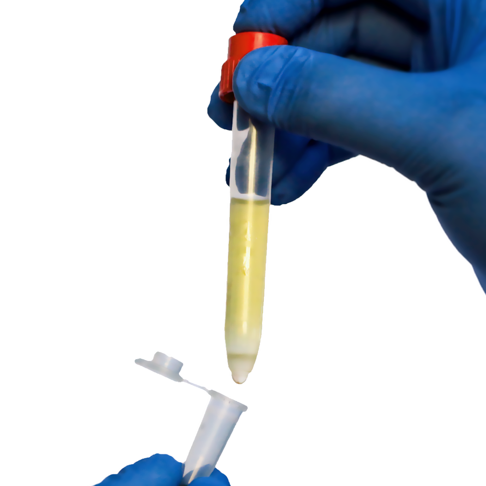
Preparing a single-cell suspension is critical for successful cell isolation, but clumpy cell samples can reduce recovery and interfere with the labeling of target cells. When samples are subjected to repeated freeze/thaw cycles or enzymatic tissue dissociation, they may appear “clumpy.” Environmental stresses can hasten the rate of cell death within the sample, resulting in the release of “sticky” DNA molecules from dying cells, which can clump neighboring cells together.
Let’s talk about one of the most serious cell culture issues: cell clumping, its causes, and how to overcome it using unique cell separation technology.
It is not uncommon to see cell loss in a sample when working with single-suspension cell cultures in a growth medium. When cells rupture, DNA and debris are released, causing cells to aggregate into large clumps that make expansion difficult. Cell clumping can both cause and lead to apoptosis or cell death.
The problem will worsen as more cells die and release the debris. Dealing with cell clumping as soon as possible or avoiding it altogether will benefit an experiment’s downstream results.
Why is Cell Clumping a Problem?
When cells clump together, they restrict each other, lowering throughput and potentially compromising downstream results. Cell clumping also reduces target cell recovery and can interfere with labeling.
Certain cell analysis methods, such as flow cytometry, necessitate proper cell isolation. Flow cytometry involves passing a large volume of cells through a machine in a rapid fluid stream.
The machine, known as a cytometer, detects physical differences between cells and labels them accordingly using fluorescent light. The cytometer will struggle to accurately measure the characteristics of clustered cells as they pass through the tube. This can lead to them being incorrectly sorted, which can have an adverse effect on downstream results.
Causes of Cell Clumping
Cells in culture may clump for a variety of reasons, including:
- Over-digestion — Excessive use of certain enzymes used for tissue dissociation, such as trypsin, can cause cells to clump.
- Environmental stress — Physical forces and repeated temperature changes can cause cell death, resulting in “sticky” DNA molecules that connect neighboring cells.
- Tissue disaggregation — When preparing a single-cell suspension, using enzymatic, mechanical, or chemical dissociation strategies can rupture cells, resulting in clumping debris.
- Overgrowth — When cells reach confluency or their maximum growth potential in their culture medium, they begin to lyse and release the debris.
- Contamination — Certain bacterial and fungal pathogens cause cells to lyse; clumping is frequently a sign that a cell culture has been contaminated.
Reducing Cell Clumping
Before beginning an experiment, precautions can be taken to prevent cells from aggregating. For example, DNase I, an endonuclease, can be mixed into a sample to fragment the DNA from ruptured cells. Breaking up this extra debris allows more room for target cells to grow. DNase I, on the other hand, should not be used when intending to engineer or change cells downstream because it can affect cell health and physiology.
Read More: CTCs: What are They and How do separate them with a high level of purity?
Cell clumps can also be reduced by using proper equipment and handling. Setting the correct speed on a centrifuge to separate or mix a sample can reduce the chances of buildup. Faster speeds typically prevent cells from piling up, but the fragility of cells must also be considered when subjecting them to high velocity.
Unclumping Cells
There are also methods for separating cell clumps without causing too much damage. Chelators are chemicals that have an affinity for positively charged ions. Chelators can be added to dissolve the bonds between the clumps without harming the cells, depending on the nature of the clumps. A chelator that is commonly used to dissolve calcium bonds is ethylenediaminetetraacetic acid (EDTA).
Trituration is an alternative method for unclumping cells. Trituration is the gentle, repetitive pipetting of a sample to break up weak cell bonds.
Pluriselect’s Innovative Technologies for Eliminating Cell Clumping
There is less residual DNA and debris floating around in the sample as a result of effectively sorting cells in a gentle manner that preserves cell health and physiology. Pluriselect’sCell Separation products offer a quick and efficient cell separation method that yields a highly enriched sample of the target cells.
The three cell isolation technologies that we have developed with efficiency in mind are listed below:
- Antibody-supported cell separation: PluriSelect provides the pluriSpin negative isolation and depletion system, as well as the pluriBead positive isolation system. The pluriSpin system isolates negative cells directly from whole blood, buffy coat, or cord blood. This new method isolates untouched, highly purified, and viable cells in a single step without the use of magnets or a column. As a result, the targeted cells are less likely to be activated or damaged. PluriBead is a one-of-a-kind cell separation technology that does not use magnetic components. The procedure is simple: the pluriBeads (containing bound target cells) are sieved through a strainer, with the pluriBeads containing your target cells remaining on top and the unwanted cells passing through. After detaching, you have your target cells ready.
- Gravity Supported Separation: PluriSelect provides optimized density gradient media for the isolation of Leukocytes, Monocytes, Platelets, and PBMCs from older blood, as well as the most commonly used solution for PBMC isolation (density 1.077 g/ml).
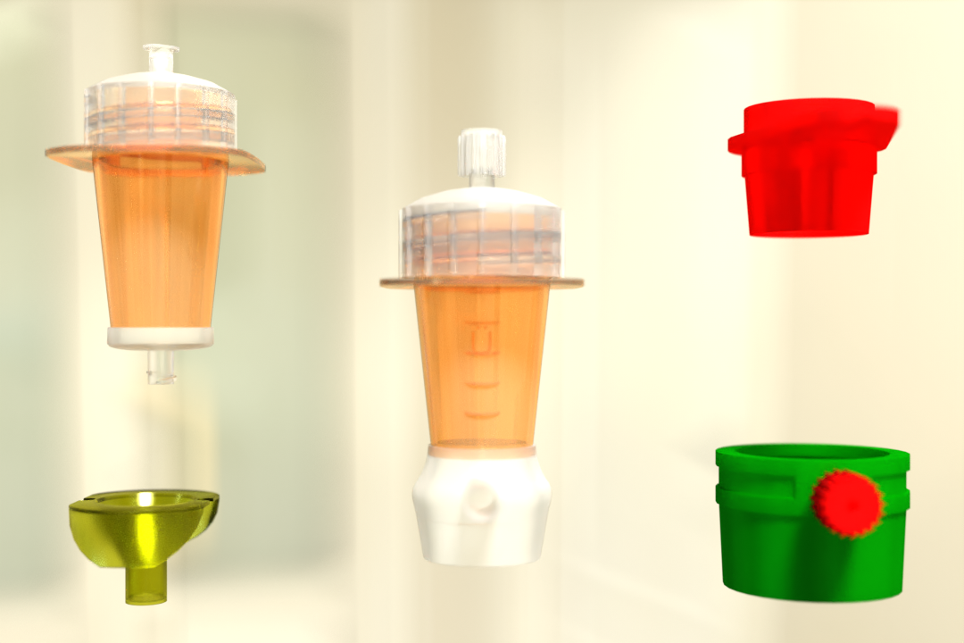
- Size Support Separation: Pluriselect Strainers are ideal for sample preparation. Choose from meh sizes ranging from 1 um to 1000 um, sterile or non-sterile, PET or nylon fibers, back-flush capability, and small volumes up to 800 uL or in-line-strainer for your sample preparation. The various straining/filtration devices available are PluriStrainer Mini, PluriStrainer, ÜberStrainer, Syringe Strainer, Membrane Strainer, Steel Basket-Strainer, and Re-Strainer (In-Line-Strainer).
For researchers needing an efficient tool for isolating specific cell types from various sample sources, we recommend trying our huge variety of cell enrichment products based on advance cell separation technology.
 English
 French
 German
 Spanish
 Belgium
 Italian
 Brazil
 Chinese Mandarin