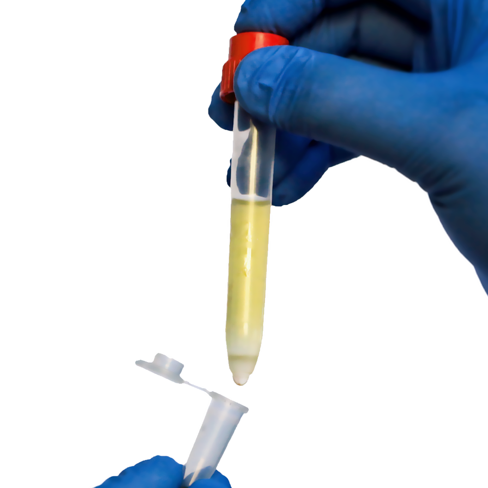
Regulatory T cells (Tregs) are a type of T cell that suppresses immune responses by secreting negative regulatory (anti-inflammatory) cytokines. Let’s find out about the Regulatory T-cells separation technologies.
T cells that regulate or suppress other immune system cells are known as regulatory T cells (also known as Tregs). Tregs help to prevent autoimmune disease by regulating the immune response to self and foreign particles (antigens). Tregs are produced naturally by a healthy thymus. ‘Adaptive’ Treg are created by differentiating naive T cells outside of the thymus, i.e. the periphery, or in cell culture.
CD4+ T cells are commonly classified as regulatory T (Treg) cells or T helper (Th) cells. The cells regulate adaptive immunity against pathogens and cancer by activating other effector immune cells. Treg cells are CD4+ T cells that are in charge of suppressing potentially harmful Th cell activities.
We’ll look at how Pluribead cascade straining aids in Regulatory T Cells isolation.
Identification of Treg cells remains difficult because evidence suggests that all of the currently used Treg markers (CD25, CTLA-4, GITR, LAG-3, CD127, and Foxp3) are general T-cell activation markers rather than Treg-specific.
Treg-cell activation is antigen-specific, implying that Treg cells’ suppressive activities are antigen-dependent. Self-reactivity of Treg cells has been proposed, but extensive TCR repertoire analysis suggests that self-reactivity may be the exception rather than the rule. Because the ability to suppress is not unique to Treg cells, their classification as a distinct lineage remains debatable. Suppressive activities attributed to Treg cells may be exerted by conventional Th cell subsets such as Th1, Th2, Th17, and T follicular cells, at least in some experimental settings.
Natural Tregs are distinguished by the expression of both the CD4 T cell co-receptor and CD25, a component of the IL-2 receptor. Tregs are therefore CD4+CD25+. The expression of the nuclear transcription factor Forkhead box P3 (FoxP3) determines natural Treg development and function.
FoxP3 is required to keep the immune system suppressed. Self-reactive lymphocytes caused by naturally occurring mutations in the FOXP3 gene cause the rare but severe disease IPEX (Immune Dysregulation, Polyendocrinopathy, Enteropathy, X-Linked) in humans and scurfy in mice.
Tregs are thought to suppress CD4+ and CD8+ T cell activation, proliferation, and cytokine production, as well as B and dendritic cells. Tregs can produce soluble messengers with suppressive properties, such as TGF-beta, IL-10, and adenosine. CD152 (CTLA-4) and GITR (glucocorticoid-induced TNF receptor) are additional markers of natural Tregs, but it should be noted that these are also expressed by other T-cell types on a regular basis (e.g. activated T cells), so they are not conclusively diagnostic. However, the function of these markers on other T cells is unclear. T cells lacking specialized regulatory capacity may compete for resources such as growth factors and MHC class II stimulation, and thus play a regulatory role through this general mechanism.
What Are The Functions Of Treg Cells?
Treg cells’ primary function was originally defined as the prevention of autoimmune diseases through self-tolerance. Several additional functions have been proposed over the years, and it will be critical to determine what Treg cells actually do in the immune system.
- Prevention of autoimmune diseases through the development and maintenance of immunologic self-tolerance.
- Allergic and asthmatic symptoms are reduced.
- Oral tolerance is the induction of tolerance against dietary antigens.
- Induction of maternal-fetal tolerance.
- Pathogen-induced immunopathology is suppressed.
- Regulation of the immune response’s effector class.
- T-cell activation is suppressed in response to weak stimuli.
- Effector T cells regulate the magnitude of the immune response.
- Protection of commensal bacteria from immune system elimination.
- T cells that have been activated by a true high-affinity agonist ligand are prevented from killing cells that only express low-affinity T-cell receptor (TCR) ligands, Like the self peptide-major histocompatibility complex (MHC) molecule, which positively selected the T cell.
How To Identify Treg Cells?
Molecular markers are critical tools for defining and analyzing immune cell subpopulations. The failure to identify specific markers for suppressor T cells contributed significantly to the decline of suppressor T cells at the end of the 1980s.
The most commonly used Treg cell markers are:
- CD25.
- cytotoxic T lymphocyte-associated antigen 4 (CTLA-4).
- TNF receptor family-related gene induced by glucocorticoids (GITR).
- lymphocyte activation gene-3 (LAG-3).
- P3 forkhead/winged-helix transcription factor box CD127 (Foxp3).
Upon activation, all T cells express CD25, the -chain of the interleukin-2 (IL-2) receptor, IL-2 is a T-cell growth factor important for T-cell clonal expansion. CTLA-4 is a negative regulator of T-cell activation that is upregulated on all CD4+ and CD8+ T cells 2-3 days after activation. Similarly, T-cell activation induces the expression of GITR and LAG-3.
It has been proposed that CD127, the IL-7 receptor chain, could be used to distinguish CD127low Treg cells from CD127high conventional Th cells in humans. However, it has recently been discovered that most CD4+ T cells downregulate CD127 when activated. Furthermore, loss of CD127 is a characteristic feature of T follicular helper (Tfh) cells in human tonsils, which help B cells.
It has been reported that when activated, naive, CD25-negative mouse CD4+ T cells do not upregulate Foxp3. However, it is now well established that most human CD4+ and CD8+ T cells express Foxp3 transiently upon activation.
Finally, all of the Treg markers currently in use (CD25, CTLA-4, GITR, CD127, LAG-3, and Foxp3) appear to be general T-cell activation markers. This finding strongly implies that T-cell activation is required for T-cell suppression. However, it also implies that current Treg markers are not truly Treg-specific and, as a result, are ineffective at distinguishing Treg cells from activated conventional Th cells.
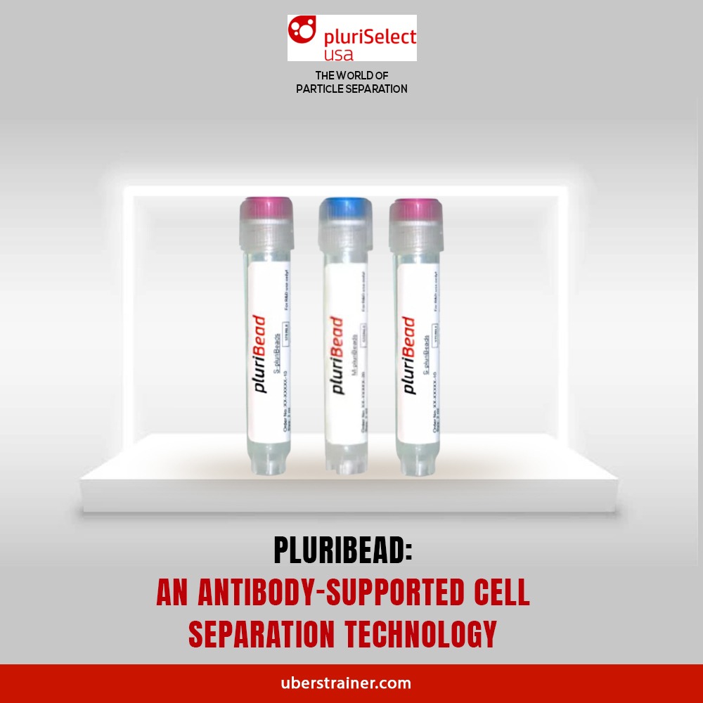
Pluribeads For Regulatory T Cells Separation
Pluribead Antibody Cell Separation helps in the gentle and safe isolation of Dendritic Cells and operates without the use of any magnetic components.
The method is simple: your pluriBeads (which contain bound target cells) are sieved through a strainer, with your target cells remaining on top and unwanted cells passing through. You are now ready to proceed with your target cells after detaching.
Key features of Pluribead
- Using a sample volume ranging from 200 l to 45 ml, no erythrolysis, gradient centrifugation, or other techniques should be used.
- Any type of sample material, such as PBMC, secretion/excretion material, liver, spleen, buffy coat, brain homogenate, whole blood, and so on, can be used.
- Isolate from a Variety of Species: Isolate from sheep, mice, rats, cows, dogs, and other animals.
- PluriBead Cascade Straining for Simultaneous Cell Isolation: Separate two different cell types from the same sample material at the same time.
- You can isolate up to six different targets from a single sample using sequential cell isolation.
Visit our website to learn more about us. You can see the difference for yourself by using our products. Order our cell separation products as soon as possible, and don’t forget to check out our Programs and Special Offers!
Reference:
Pubmed
Nature
 English
 French
 German
 Spanish
 Belgium
 Italian
 Brazil
 Chinese Mandarin


