Antigens are molecules found on the surface of cells that bind to receptors found on antibodies or lymphocytes. Antigens are classified according to their origin, and the immune system distinguishes between native and foreign antigens in order to fight pathogens. Immunology research makes use of antigen-antibody binding properties to detect, track, and ensure cell enrichment of specific cell populations within blood samples.
Self-Antigens vs Non-Self Antigens
Antigens are classified into three types based on their origin. Autoantigens are produced within the body’s cells; endogenous antigens are produced within the body by bacteria or viruses, and exogenous antigens are produced outside the body and are foreign to the immune system. Autoantigens are referred to as “self-antigens,” whereas endogenous and exogenous antigens are referred to as “non-self antigens.”
Self-Antigen Examples
Self-antigens found on the surface of leucocytes (including lymphocytes such as B and T cells) are referred to as “clusters of differentiation.” A differentiation cluster [CD] on a B or T cell can function as a cellular marker that identifies the cell to the rest of the body. Differentiation clusters can also act as receptors or ligands (activating receptors on other cells) to aid in cell signaling, allowing the cell expressing the CD to influence or manipulate the behavior of other cells in the body. CDs also play a role in the adaptive immune response, signaling B cells to produce antibodies that can bind to specific non-self antigens on pathogen surfaces, or signaling T cells or other macrophages to coordinate attacks on hostile foreign cells.
The antibodies or cellular receptors to which clusters of differentiation self-antigens bind are used to describe them. Immunologists frequently use a collection of differentiation lists to identify lymphocytes and the roles they play in the immune system. Helper T cells, which release cell-signaling cytokines when activated, are referred to as “CD4+ cells,” whereas killer T cells, which attack pathogens directly by secreting cytotoxins, are referred to as “CD8+ cells.”
During the development of B and T cells, the body ensures that self-antigens do not elicit an immune response through a process known as “central tolerance.” During cellular development, lymphocytes with antigen-binding sites that may bind to and become activated by self-antigens are eliminated through central tolerance, also known as “negative selection.” Only lymphocytes that are unreactive to self-antigens (that is, only B or T cells that react to non-self-antigens) reach maturity and participate in the immune system in this manner.
Examples of Non-Self Antigens
Antigen structures found on the surfaces of foreign bacteria, viruses, fungi, parasites, or other non-native bio compounds are examples of non-self-antigens. To recognize non-self-antigens within the body, the immune system relies on antibody-antigen binding and antigen-binding to receptors on B or T cells. Non-self-antigen binding to antibodies or lymphocytes frequently triggers an immune response, which can result in the neutralization of the pathogenic cell presenting the non-self-antigen or flagging of the non-self-antigens for recognition by other immune system components.
Antigens in Immunology Research
Scientists can introduce antibodies capable of binding to specific clusters of differentiation in order to track or manipulate their behavior, similar to how the immune system uses antibodies to flag non-self-antigens on the surface of foreign cells. We will go over the innovative Antibody Cell Separation technologies in detail below.
Antigen-based vaccines, for example, train the immune system to fight pathogens by exposing a patient to specific non-self antigens in order to activate B cells to produce antibodies that can fight or flag the pathogen in question in the event of future infection. In a research setting, specific cells within a heterogeneous cell population can be targeted, identified, marked, and even separated from the rest of the sample using the exclusive binding properties of the antibody-antigen coupling.
Try Pluriselect’s Cell Separation Products Today
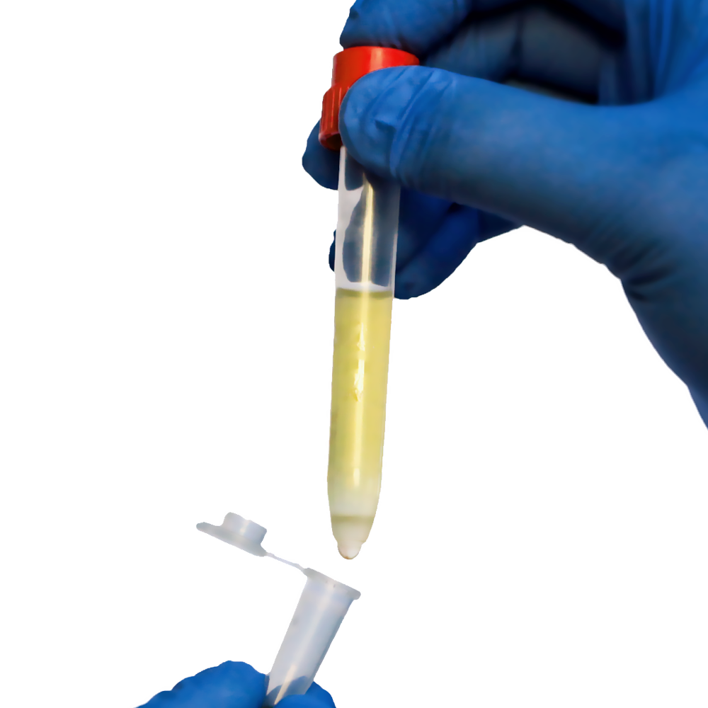
If you’re looking for the most efficient way to separate T cells from unwanted cell populations, pluriBead and pluriSpin technology provides an exceptionally gentle method for T cell enrichment that preserves the physiology and health of delicate immune cells.
Plurispin
Negative cell selection eliminates all unwanted cells. In contrast to positive cell enrichment, all cells, with the exception of the cells of interest, will be bound to specific antibodies and separated. While the unwanted cells are depleted, the desired cells remain unbound and “untouched” by antibodies or beads in the sample material.
PluriSelect’spluriSpin system isolates viable, untouched, and highly purified cells in a single step without the use of magnets or columns. As a result, the targeted cells are less likely to be damaged or activated.
PluriSpin requires no specialized training or equipment, such as special instruments or magnets. Only a mixer, such as a whipping rolling mixer or our magnetic stirrer adapter, is required (pluriPlix).
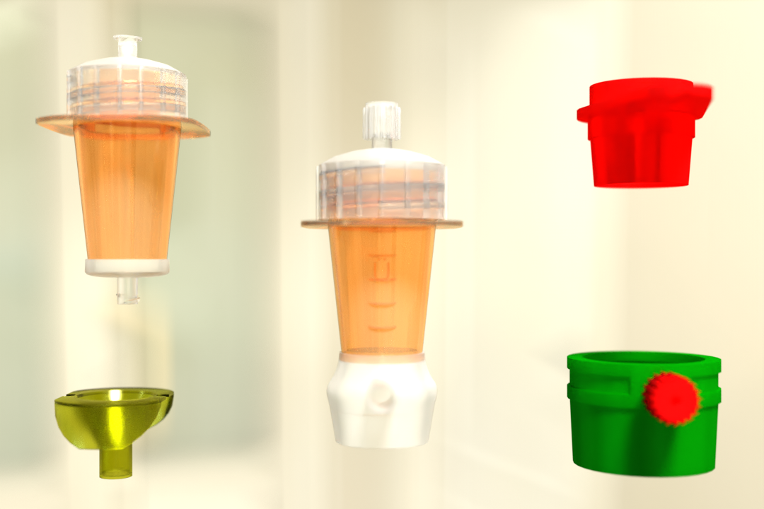
PluriBead
A specific antibody binds directly to the cells using positive selection.The cells will come into contact with the antibody.During the enrichment steps that follow, all unwanted cells remain unbound and will be separated from the labeled and desired cells.
Read Also: What Is PBMC? Human PBMC Cells, PBMC Composition, and Isolation Tools
When coupled to a solid phase, the simplest way to keep labeled cells in place is to use a cell strainer or magnets. Any sample material, such as whole blood, buffy coat, PBMC, secretion/excretion material, brain homogenate, spleen, liver, and so on, can be used. It can be used on a wide variety of species, including mice, rats, bovines, humans, canines, sheep, and others.
There are two bead sizes available: S-pluriBead, which is recommended for a small number of targets in a large sample volume (e.g. CTC), and M-pluriBead, which is recommended for a large number of targets in small materials (e.g. buffy coat).
While traditional cell enrichment methods subject samples to intense mechanical or magnetic forces and necessitate the use of expensive equipment, Pluriselect’s Cell Separation products are gentle, cost-effective, and simple to use. Furthermore, the techniques provide accurate, scalable cell isolation for both small labs and large research institutions. Learn more about how to isolate T cells with pluribead and plurispin to study pathogens with non-self-antigens on our products page.
Reference:
Frontiersin
Cell
 English
 French
 German
 Spanish
 Belgium
 Italian
 Brazil
 Chinese Mandarin