Schwann cells surround neurons, keeping them alive and occasionally encasing them in a myelin sheath. To learn more about Schwann cells, keep reading.
The neural crest is the embryological source of Schwann cells. They act as the primary glial cells of the peripheral nervous system (PNS), myelinating peripheral nerves and supplying axons with nutrients and insulation.
Conduction velocity along the axon is accelerated by myelination, enabling saltatory conduction of impulses. Nonmyelinating Schwann cells still offer trophic support and cushioning to the unmyelinated axons even though they do not wrap axons to improve conduction.
Structure of Schwann Cells
Numerous Schwann cells are required to myelinate the length of an axon because each Schwann cell contributes to one myelin sheath on a peripheral axon, and a different Schwann cell creates each subsequent myelin sheath. The central nervous system’s (CNS) myelinating cell, the oligodendrocyte, forms myelin sheaths for numerous surrounding axons, in contrast to this arrangement.
Unlike oligodendrocytes, Schwann cells are surrounded by a basal lamina. Nodes of Ranvier, or gaps of about 1 micrometer, exist between adjacent myelin sheaths. The node, the site of saltatory conduction, contains a concentration of voltage-gated sodium channels. In densely myelinated neurons, Schmidt-Lanterman incisures are cytoplasmic outpouchings that disrupt compact myelin. They have a high density of gap junctions and other cell junctions, which help the Schwann cell communicate and stay healthy.
The Function of Schwann Cells
The PNS’s myelinating cell and the peripheral neurons’ support cells are both Schwann cells. By concentrically wrapping its plasma membrane around the inner axon, a Schwann cell creates a myelin sheath. The inner turn of the glial cell membrane spirals around the axon to add membrane layers, or lamellae, to the myelin sheath while the nucleus stays fixed. Extremely high levels of lipids can be found in the plasma membrane of Schwann cells, and cholesterol plays a crucial role in the construction of the myelin sheath.
The axon segment is protected by the compact myelin sheath, which also increases conduction velocity and significantly lowers membrane capacitance. The survival and maturation of Schwann cell precursors depend on neuregulin-type III expression on axons, and the amount of neuregulin present on the axon surface determines the degree of myelination.
Axons are also given energy metabolites by Schwann cells, which shuttle them through monocarboxylate transports that are located on the axon’s surface and inside the Schwann cell. In response to PNS axon damage and axon regeneration, Schwann cells are essential. Wallerian degeneration will happen away from the area of injury.
When the distal axon segment dies, Schwann cells and then macrophages clear the contents of the dead cells and encourage axon regeneration. At this time, Schwann cells go through a number of phenotypic changes: they activate the breakdown of myelin, up-regulate the expression of cytokines (such as TNF-a), which attract macrophages to the site of the injury, up-regulate neurotrophic factors, which promote axon regeneration and neuron survival, and organize a regeneration pathway along their basal lamina tube, which directs axon growth.
The two main types of PNS nerve injury are axonotmesis and neurotmesis. The axon is damaged in axonotmesis, such as in a crush injury, but the Schwann cells’ basal lamina tube is still present. The regenerating axon sprout receives growth-guiding cues from the tube lumen, which accelerates axon regeneration and restores function in 3 to 4 weeks. The axon, Schwann cell basal lamina, and surrounding connective tissue sheath is damaged during neurotmesis, such as during a cut injury.
From the proximal to the distal nerve stump, the regenerating axon and the Schwann cells that are related to it continue to grow. In the absence of the basal lamina tube, targeting errors make it difficult to reinnervate correctly and restore function in neurotmesis.
Schwann cells and axon regeneration
In order to regenerate axons, Schwann cells are essential. Axonal degeneration and cell death are possible outcomes of any axonal injury. Upon injury, macrophages and Schwann cells are drawn to the wound site to clear away dead tissue and encourage axonal regeneration. Myelin breakdown and increased expression of cytokines like tumor necrosis factor-alpha are two mechanisms by which Schwann cells encourage the recruitment of macrophages.
Additionally, Schwann cells promote the expression of a wide range of growth factors, including neurotrophins, transforming growth factor-beta, glial cell line-derived neurotrophic factor, epidermal growth factors, and platelet-derived growth factor, to promote axon regeneration and nerve cell survival. To aid in the regeneration process, they also secrete extracellular matrix molecules like laminin and collagen as well as cell adhesion molecules. The cues given by the Schwann cell’s basal lamina lumen serve to direct axonal growth.
Pluribeads: an innovative approach to cell separation technology
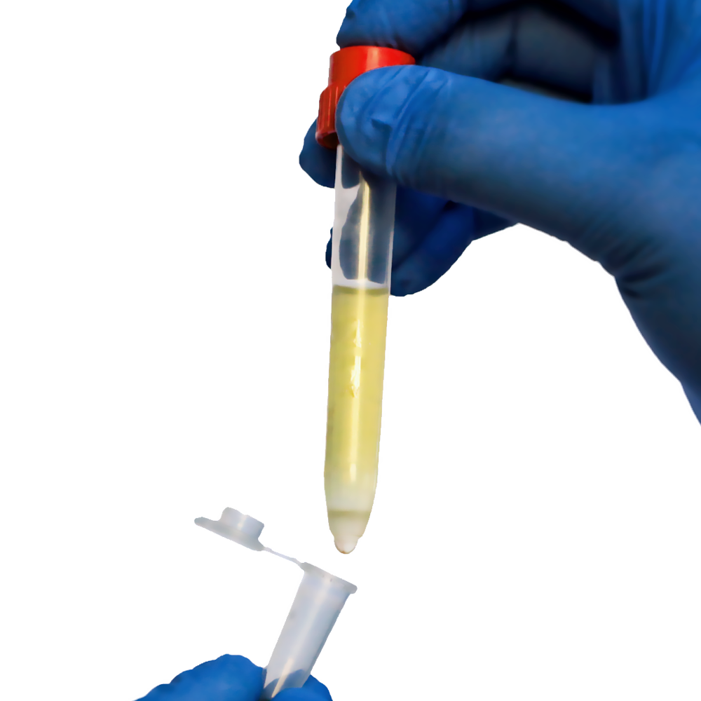
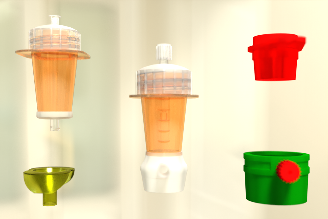
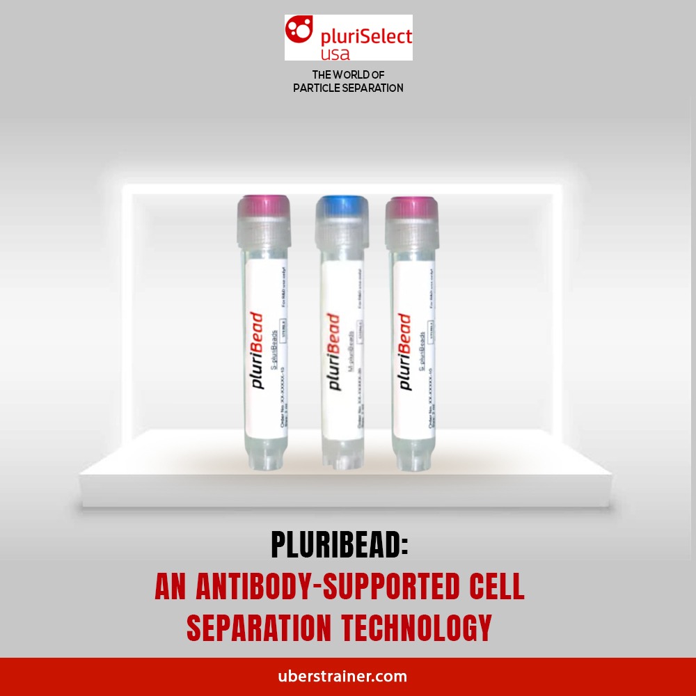
Pluribead is one such Cell Separation technology that helps in the gentle and safe isolation of Schwann cells. Unusual cell separation technology called PluriBead operates devoid of any magnetic components. The process is easy: Your pluriBeads (which contain bound target cells) are sieved through a strainer; the pluriBeads containing your target cells remain on top while the unwanted cells pass through. You are prepared with your target cells after detaching.
Key features of Pluribead
- No Sample Preparation: Utilize a sample volume of 200 l to 45 ml without the use of erythrolysis, gradient centrifugation, or any other techniques.
- Use any type of sample material, including PBMC, secretion/excretion material, brain homogenate, spleen, liver, buffy coat, whole blood, and so on.
- Applicable to a Wide Variety of Species: Isolate from sheep, mice, rats, cows, dogs, and other animals.
- Quick Isolation: Cell division can begin in five minutes.
- Simultaneous Cell Isolation Using PluriBead Cascade: Separate two different cell types simultaneously from the same sample material.
- Sequential Cell separation: Utilizing sequential cell isolation, isolate up to six different targets from a single sample.
Two Different Bead Sizes Available
- S-pluriBead: It is used for a small number of targets in a large sample volume.
- M-pluriBead: A versatile material that can be used for many targets while using less material (e.g. buffy coat).
You can find out more about us by going to our website. By using our products, you can see the difference for yourself. Begin using our cell separation products right away!
Reference:
Science Direct
Britannica
 English
 French
 German
 Spanish
 Belgium
 Italian
 Brazil
 Chinese Mandarin
