A buffy coat contains a concentrated suspension of leukocytes derived from bone marrow or whole blood. This blog discusses how to prepare the buffy coat.
White blood cells (WBCs) and platelets are concentrated in a buffy coat. WBCs and platelets account for only about 1% of the cells in peripheral whole blood. When a blood sample undergoes density gradient centrifugation, the platelets and WBCs unite to form a layer floating between the RBCs and supernatant plasma. This thin coating is known as a buffy coat because of its color.
This blog will look at the different cell types found in buffy coats, how buffy coats are extracted from whole blood and the significance of this material for research.
Four Cell Types Exist in Buffy Coats
- Granulocytes are immune cells such as basophils, eosinophils, and neutrophils.
- Platelets, also known as thrombocytes, play various roles in blood homeostasis.
- Lymphocytes – Lymphocytes play an important role in the immune system. T cells, B cells, and natural killer cells are among them.
- Monocytes- They are leukocytes that help the immune system by identifying and digesting microbes. Monocytes have the ability to differentiate into dendritic cells or macrophages.
The cells found in a buffy coat play an important role in the pathogenesis of the human disease. The study of buffy coat cells benefits research and provides valuable scientific information.
Is the Buffy Coat the Same as PBMCs?
Buffy coat and peripheral blood mononuclear cells (PBMCs) are terms that are frequently used interchangeably. They are not, however, the same, and they serve different functions in research.
PBMCs contain lymphocytes and monocytes, which remain a subset of all immune cells and do not include other buffy coat cell types such as eosinophils, basophils, and neutrophils. One of several density centrifugation techniques is used to isolate PBMCs from whole blood. Centrifugation causes the relatively lower-density PBMCs to migrate into a layer that floats on top of the reagent layer, where they can be easily extracted.
What Is the Purpose of Buffy Coats in Research?
Buffy coats are high in WBCs and platelets, making them ideal for studying immunological pathways and molecular signals.
Buffy coats are commonly used to extract DNA from mammalian blood. A small amount of buffy-coat DNA can yield a large amount of DNA with high quality, integrity, and functionality. Cell pellets derived from buffy coats can be stored at -80 °C for up to 9 years and still yield high yields of DNA suitable for whole-genome sequencing, genome-wide association analysis, and another genetic testing.
Circulating tumor cells (CTCs) can be found in the buffy coats of cancer patients and can aid in the downstream analysis of potentially critical tumor molecular mechanisms.
Whole blood samples are frequently separated in blood banks in order to extract the buffy coat and cryopreserve platelets for future use.
The buffy coat can be useful in diagnostics. Because important immune cells congregate in this layer, it can be used to detect parasitic infections such as plasmodium, microfilariae, and Trypanosoma. Buffy coats are an important starting material for immunology research as well as other cell enrichment applications.
Buffy Coat vs. PBMC
The terms buffy coat and PBMC are frequently used interchangeably; however, minor differences in cell composition can make a significant difference. A buffy coat is a centrifuged mixture of lymphocytes, monocytes, granulocytes, and platelets from plasma and RBCs. Individually fragmented lymphocytes and monocytes, on the other hand, separate from the rest of the whole blood sample using a technique called density-gradient centrifugation.
In this process, an additional density gradient is used to further separate sample particles based on their size and density. By removing granulocytes and platelets and forming a barrier between WBCs and RBCs, the sample becomes more concentrated in monocytes and lymphocytes, which can then be used for research or isolated for specific downstream applications.
Buffy Coat Extraction
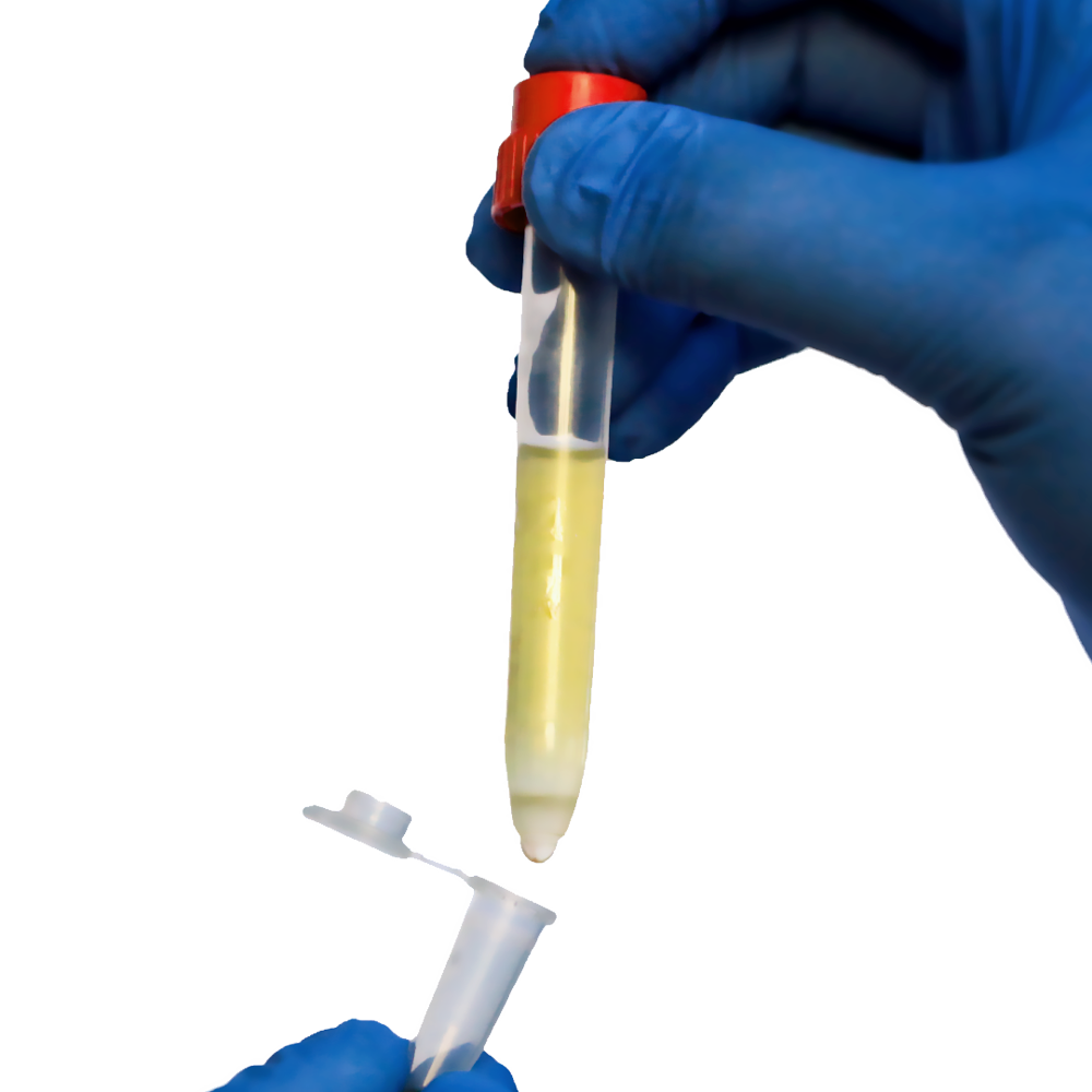
The buffy coat is separated from the plasma and RBC in a centrifuge after the prepared whole blood sample has been spun. The buffy coat is a thin layer between the RBC and plasma that accounts for about 1% of the sorted sample after centrifugation. Using a small pipette, the experimenter collects the buffy coat and transfers it to a separate container.
The buffy coat is in direct contact with isolated RBCs during PBMC isolation, which frequently involves particle separation by a density gradient in the sorted sample. Because of the size and abundance of the RBCs, using a manual extraction method like pipetting increases the likelihood that contaminating RBCs are included in the buffy coat sample (Prior to further processing, buffy coats are referred to as “pink” because of this). Researchers will frequently combine centrifugation with another method of cell separation, such as BACS, to remove residual RBC contamination in order to enrich the buffy coat even more.
How to Prepare a Buffy Coat fraction out of fresh whole blood in your lab?
Another option is to make your own buffy coat. Simply follow the following short protocol:
- Mix 1 part of whole blood with one part of washing buffer
- With the help of the density gradient centrifugation technique, the diluted whole blood is centrifuged for 10 Minutes at 200 x g with the brake off
- Remove the interphase leukocytes (buffy coat)
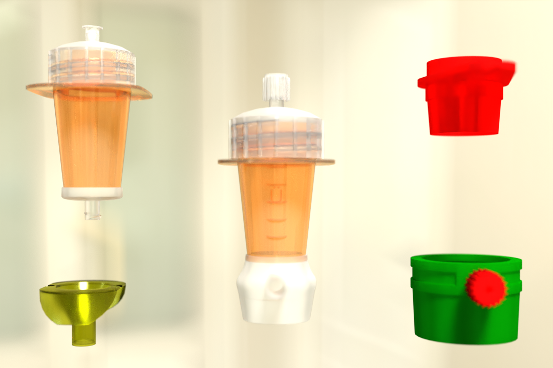
Pluribead’s Cell Separation Technology
Pluribead is an innovative technology developed by Pluriselect that helps in the gentle and safe isolation of cells. The procedure is simple and clear: your pluriBeads (which contain bound target cells) are sieved through a strainer, with the pluriBeads containing your target cells remaining on top while unwanted cells pass through.
After detaching, you are ready to go with your target cells. Traditional blood separation techniques necessitate costly equipment, lengthy processing times, and unneeded cell death. Pluribead, on the other hand, allows for a simplified workflow with no additional equipment and sample enrichment that is exceptionally gentle on cells.
Read More About: Pluriselect’s All-Star-Strainer-Team: One Stop Solution to Particle Separation
Do you want to learn more about buffy coats or request a specific biospecimen? Then contact us to see how we can help you with your research! Also, don’t forget to check out the Programs & Special offer section on our website.
Reference:
Science Direct
Parasites And Vectors
NCBI
 English
 French
 German
 Spanish
 Belgium
 Italian
 Brazil
 Chinese Mandarin