Buffy coat preparation is frequently the first step in further processing for T-cell depletion, tumor cell purging, or cryopreservation of both marrow and blood HPC collections.
White blood cells (WBCs) and platelets make up less than 1% of the cells in a sample of peripheral whole blood. When researchers spin the blood sample in a centrifuge, the WBCs and platelets combine to form their own layer suspended between the red blood cells (RBCs) and supernatant plasma. Because of its color, this thin layer is known as a buffy coat (yellowish to brownish). The formation of a buffy coat from whole blood samples aids in the concentration of large sample volumes and the reduction of downstream cell separation handling.
The buffy coat contains 10-20X more leukocytes (white blood cells that help the body fight infection) than the rest of the sample. This can be useful for toxicology researchers or patients with dysfunctional WBCs or platelets.
Buffy Coat Cells
A buffy coat is made up of four different types of cells:
- Lymphocytes – A type of WBC that produces antibodies and thus plays an important role in the human immune system. Lymphocytes are further subdivided into T cells, B cells, and natural killer cells.
- Monocytes- These are a type of white blood cell that aids in the immune response by isolating and digesting harmful microorganisms. Monocytes can differentiate into either macrophages or dendritic cells.
- Granulocytes – A type of immune cell that is further subdivided into basophils, eosinophils, and neutrophils, which secrete chemicals, attack parasites, and kill bacteria/fungi, in that order.
- Platelets – These cells, also known as thrombocytes, are not RBCs or WBCs. They are small colorless cell fragments that migrate throughout the body, forming clots and preventing bleeding as needed.
The cells found in a buffy coat are critical in protecting humans from disease and healing wounds.
Importance of a Buffy Coat
White blood cells serve as the foundation of the human immune system. Different types collaborate to detect foreign substances, eliminate threats to the body, and establish a long-term defense system against future infections. Platelets are the human body’s first responders, rushing to injured areas and clotting blood flow through adhesion. WBCs and platelets are both present and abundant in the buffy coat. The buffy coat is a critical bio-fluid in the medical research field due to its high concentration of WBCs and platelets, allowing academic professionals to conduct experiments and study these essential cells and how they work in the body — which can shape the direction of medicine and effective patient treatment.
Aside from its research implications, the buffy coat has a practical application in disease testing and treatment. Because key immune cells congregate in this layer, the buffy coat can be used to screen for the presence of dangerous diseases such as malaria. Platelets can also be purified further and used therapeutically to boost counts in patients with low platelet counts.
Buffy Coat vs. PBMC
Although the terms buffy coat and PBMC are frequently used interchangeably, there are minor differences in cell composition that can make a significant difference. A buffy coat is a mixture of lymphocytes, monocytes, granulocytes, and platelets that have been centrifuged from plasma and RBCs. Individual fragmented lymphocytes and monocytes, on the other hand, separate from the rest of the whole blood sample via a process known as density-gradient centrifugation.
An additional density gradient is used in this process to further separate sample particles based on their size and density. By removing granulocytes and platelets and forming a barrier between the WBCs and the RBCs, the sample becomes more concentrated in monocytes and lymphocytes, which can then be used for research purposes, or isolated further for specific downstream applications.
How to Prepare a Buffy Coat
Although the contents of a buffy coat exist in whole blood, they are not aggregated until centrifugation occurs. Researchers should take a number of precautions prior to centrifugation to ensure the extraction process is successful.
There are two methods for preparing a buffy coat. On one hand, whole blood donations are centrifuged to separate red cells, plasma, and buffy coats. The second option is to filter the blood and keep the leukocytes at bay.
How to Prepare a Buffy Coat fraction out of fresh whole blood?
Another option is to make the buffy coat from scratch. As a result, simply follow the following short protocol:
Mix 1 part whole blood to 1 part washing buffer
Centrifuge the diluted whole blood for 10 minutes at 200 x g while the brake is turned off.
Remove the interphase leukocytes (buffy coat)
It’s important to remember that a whole blood sample could contain pathogens, so these samples must be handled with the same care as if they were capable of transmitting infectious diseases. After making the necessary preparations, it’s time to extract the buffy coat from a whole blood sample.
Read Also: A Detailed Guide on Positive Selection vs Negative Selection for Cell Isolation
Buffy Coat Extraction
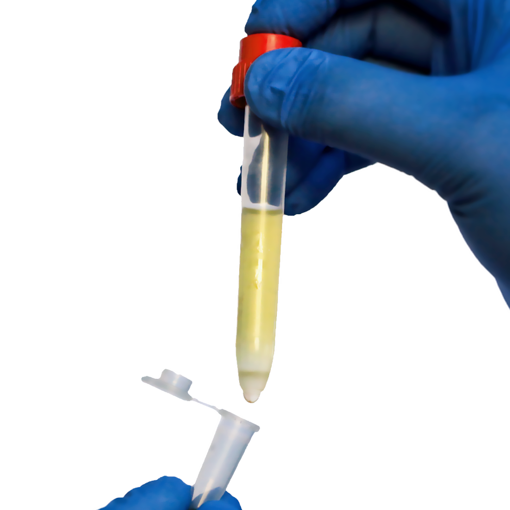
The prepared whole blood sample is spun in a centrifuge to separate the buffy coat from the plasma and RBC. After centrifugation, there will be a thin layer between the RBC and plasma that accounts for about 1% of the sorted sample – the buffy coat. The buffy coat is collected and transferred to a separate container by the experimenter using a small pipette.
While PBMCs are separated from the other particles in the sorted sample by density gradient centrifugation, the buffy coat is in direct contact with isolated RBCs. Because of the size and abundance of RBCs, using a manual extraction method like pipetting increases the likelihood that contaminating RBCs will be included in the buffy coat sample (this is why buffy coats are referred to as “pink” prior to further processing). To enrich the buffy coat, even more, researchers will frequently combine centrifugation with another method of cell separation, such as BACS, to remove residual RBC contamination.
How Plurispin Can Help?
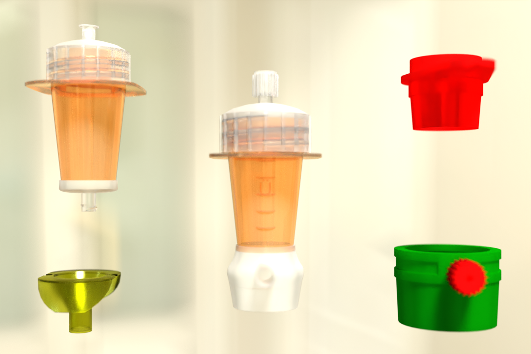
The pluriSpin system is a new negative cell isolation technology directly from whole blood, buffy coat, or cord blood. Without the use of magnets or a column, this new method isolates viable, untouched, and highly purified cells in a single step. As a result, the chances of activating or damaging the cells of interest are reduced.
How PluriBead Can Help?
pluriBead is a one-of-a-kind cell separation technology that doesn’t rely on magnetic components. The procedure is straightforward: Your pluriBeads (with bound target cells) are sieved through a strainer; the pluriBeads with your target cells remain on top, while the unwanted cells pass through. After detaching, you have your target cells ready.
For more information, you can visit our website. Experience the difference for yourself by using our products. Get started with our cell separation products today!
Reference
Science Direct
Parasites And Vectors
NCBI
 English
 French
 German
 Spanish
 Belgium
 Italian
 Brazil
 Chinese Mandarin